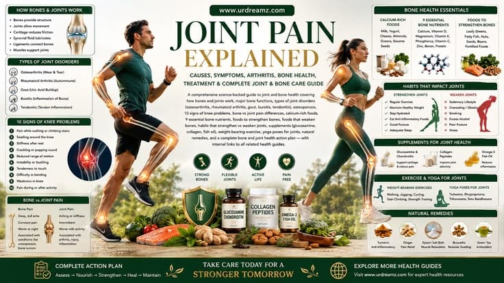
Joint Pain Explained: Causes, Symptoms, Arthritis, Bone Health, Treatment and Complete Joint & Bone Care Guide
Your bones and joints form the structural foundation of your entire physical existence. Your 206 bones — forming the skeleton that houses your organs, anchors your muscles, manufactures your blood cells, stores vital minerals, and enables every movement you make — work in seamless partnership with the cartilage, ligaments, tendons, synovial fluid, and muscles that constitute your joints. When this musculoskeletal system functions well, you move freely, stand upright, carry weight, and live actively without a second thought. When it breaks down — through arthritis, osteoporosis, injury, nutritional deficiency, or the cumulative wear of years — the consequences affect every aspect of daily life with pain, stiffness, reduced mobility, and loss of independence. Joint and bone disease is the leading cause of physical disability worldwide, affecting more than 1.7 billion people globally. This comprehensive guide covers everything you need to know about joint and bone health: how your skeleton is built and maintained, the full spectrum of bone and joint disorders, the nutrients and foods your skeleton needs, the exercises that build bone density and joint resilience, natural support strategies, and a complete plan for lifelong musculoskeletal health.
1. The Human Skeleton: Major Bones and Their Functions
The adult human skeleton consists of 206 bones — a number that begins at approximately 270 in newborns, with many bones gradually fusing throughout childhood and adolescence until the adult count is reached in the early 20s. Far from being the inert, lifeless framework that preserved skeletons in museums suggest, living bone is a dynamic, metabolically active tissue that is continuously being broken down and rebuilt, responds to mechanical forces and hormonal signals, manufactures blood cells, stores essential minerals, and performs functions without which survival would be impossible. Understanding the major bones of the skeleton and their specific functions provides the anatomical foundation for understanding what goes wrong when joints and bones are diseased or injured.
| Bone / Region | Location | Primary Functions | Common Problems When Affected |
|---|---|---|---|
| Cranium (Skull) | Head | Protects the brain; houses sensory organs (eyes, ears, nose); forms the facial structure | Skull fractures; intracranial pressure; temporal bone involvement in hearing loss |
| Mandible (Jawbone) | Lower face | Enables chewing, biting, and speaking; provides facial structure; anchors lower teeth | Temporomandibular joint (TMJ) disorder; mandibular fractures; dental disease |
| Clavicle (Collarbone) | Upper chest, connecting shoulder to sternum | Connects the arm to the axial skeleton; provides attachment for shoulder and chest muscles; protects underlying neurovascular structures | One of the most commonly fractured bones (often from falls on outstretched hand or direct impact) |
| Scapula (Shoulder Blade) | Upper back, behind the ribs | Provides attachment points for 17 muscles; enables a wide range of shoulder movement; forms the glenoid cavity of the shoulder joint | Scapular winging (serratus anterior weakness); impingement syndrome; rotator cuff problems |
| Sternum (Breastbone) | Center of the chest | Protects the heart, lungs, and major blood vessels; serves as attachment point for ribs and clavicles; site of red marrow for blood cell production | Costochondritis; sternal fractures; sternotomy in cardiac surgery |
| Ribs and Rib Cage | Encircling the chest | Protect the heart and lungs; assist in breathing mechanics through expansion and contraction; provide attachment for respiratory muscles | Rib fractures; costochondritis; pleuritis involving the parietal pleura attached to ribs |
| Vertebral Column (Spine) | Running from skull base to pelvis | Protects the spinal cord; supports the upright posture; provides attachment for ribs and muscles; enables flexible movement; transmits body weight to the pelvis | Intervertebral disc herniation; spondylosis; osteoporotic compression fractures; scoliosis; spinal stenosis |
| Humerus (Upper Arm) | Between shoulder and elbow | Connects the shoulder joint to the elbow; provides attachment for arm muscles (biceps, triceps, deltoid); forms the ball of the glenohumeral joint | Proximal humerus fractures (common in osteoporotic elderly); rotator cuff tears; bicipital tendinopathy |
| Radius and Ulna (Forearm) | Between elbow and wrist | Allow forearm rotation (pronation and supination); enable wrist and elbow movement; provide attachment for forearm muscles | Colles' fracture of radius (most common osteoporotic wrist fracture); ulnar nerve entrapment |
| Carpals and Metacarpals (Wrist and Hand) | Wrist and hand | Enable precise wrist positioning and hand movement; provide the mechanical basis for grasping, pinching, and fine motor skills | Carpal tunnel syndrome; rheumatoid arthritis (commonly affects hands early); scaphoid fractures; De Quervain's tenosynovitis |
| Pelvis (Hip Bone) | Central base of the trunk | Transfers body weight from spine to legs; protects reproductive and digestive organs; attachment point for powerful hip and core muscles; houses the hip joints | Pelvic fractures (often in osteoporotic elderly from falls); sacroiliac joint dysfunction; hip dysplasia |
| Femur (Thigh Bone) | Between hip and knee | The longest and strongest bone in the body; bears the full weight of the upper body during standing and walking; the ball of the ball-and-socket hip joint | Hip fractures (the most serious osteoporotic fracture — associated with 30% one-year mortality in elderly); femoral stress fractures in runners; avascular necrosis of the femoral head |
| Patella (Kneecap) | Front of the knee joint | Protects the knee joint from direct impact; acts as a lever increasing the mechanical advantage of the quadriceps muscle | Patellofemoral pain syndrome (runner's knee); patellar tendinopathy; patellar fractures and dislocations |
| Tibia and Fibula (Shin Bones) | Between knee and ankle | Tibia is the primary weight-bearing bone of the lower leg; fibula provides stability and muscle attachment; together they form the ankle mortise | Tibial stress fractures (common in runners — "shin splints" at extreme); ankle fractures; anterior cruciate ligament attachment at tibia |
| Tarsals and Metatarsals (Ankle and Foot) | Ankle and foot | Form the arched structure of the foot that absorbs shock during walking and running; enable ankle movement and balance; the calcaneus (heel bone) is the largest tarsal | Plantar fasciitis; ankle sprains; stress fractures in metatarsals; gout commonly affects the first metatarsophalangeal joint (big toe) |
2. How Bone Is Built and Maintained: Living Architecture
Bone is one of nature's most elegant engineering achievements — a composite material that achieves the seemingly contradictory goals of being simultaneously strong, lightweight, and flexible while also being metabolically dynamic and self-repairing. Understanding how bone is built explains both why adequate nutrition and mechanical loading are essential for bone health and why bone disease — whether osteoporosis, fractures, or metabolic bone disorders — occurs when these requirements are not met.
Bone derives its remarkable mechanical properties from two complementary components that work together like reinforced concrete. Collagen fibers — the protein framework of bone — provide flexibility and tensile strength (resistance to being pulled apart or bent), preventing the brittle cracking that would occur with mineral alone. Hydroxyapatite — a calcium phosphate mineral crystal — provides hardness, stiffness, and compressive strength (resistance to being crushed), preventing the rubber-like deformation that would occur with collagen alone. Together, these two components create a composite material far superior to either component alone — explaining why bone can absorb significant impact energy during a fall without fracturing in healthy individuals.
Bone exists in two architectural forms with complementary roles. Cortical (compact) bone forms the dense outer shell of all bones — providing rigidity, resistance to bending and twisting, and the hollow tube geometry that places the strongest material farthest from the neutral axis (maximizing bending resistance for minimum material weight — the same principle used in engineering I-beams and hollow tubes). Trabecular (cancellous or spongy) bone forms the porous internal lattice found at the ends of long bones and within flat bones — distributing complex compressive loads across a three-dimensional network, containing red marrow for blood cell production, and providing metabolically active bone surface for rapid mineral exchange. The ratio of cortical to trabecular bone varies by location: the femoral shaft is predominantly cortical (for weight-bearing and bending resistance); the femoral head and vertebral bodies are predominantly trabecular (for multidirectional load distribution).
Bone is continuously being remodeled throughout life — approximately 10% of the adult skeleton is replaced each year through a tightly regulated process of coupled bone resorption (by osteoclasts — cells that dissolve bone mineral and matrix) and bone formation (by osteoblasts — cells that synthesize new collagen and mineralize it with hydroxyapatite). This remodeling process repairs microscopic damage from daily mechanical loading, adapts bone architecture to changing load patterns (Wolff's Law — bone grows stronger in response to the loads placed upon it), and releases or deposits minerals in response to body needs. When resorption exceeds formation — due to aging, hormonal changes (particularly estrogen decline in menopause), nutritional deficiency, inactivity, or disease — bone mass declines and osteoporosis develops. The balance between osteoblast and osteoclast activity is regulated by hormones (estrogen, testosterone, PTH, calcitonin, thyroid hormones — see our Thyroid Health Guide), vitamins D and K, mechanical loading, and the coordinating actions of bone cells themselves through signaling molecules including RANKL and OPG.
3. How Joints Work: Anatomy and Function
A joint is any point where two bones meet — and joints exist on a continuum from completely immobile (the synarthrodial joints between skull bones that fuse in adulthood) to freely mobile (the diarthrodial or synovial joints that allow the wide-ranging movements of the shoulder, hip, knee, and wrist). The synovial joints — the most clinically significant type because they are the primary target of arthritis and other joint diseases — have a sophisticated architecture designed to enable smooth, low-friction movement while bearing significant mechanical loads throughout a lifetime of use.
The key structural components of a synovial joint include: articular cartilage — a layer of smooth, resilient hyaline cartilage covering the ends of the articulating bones, providing a nearly frictionless bearing surface and absorbing compressive loads through its high water content and proteoglycan matrix; the synovial membrane (synovium) — a thin, highly vascular membrane lining the inner surface of the joint capsule (excluding the articular cartilage itself), producing synovial fluid and providing nutrients to the avascular cartilage; synovial fluid — a viscous, lubricating fluid containing hyaluronic acid that reduces friction between cartilage surfaces during movement (healthy synovial joints have a friction coefficient lower than ice on ice), provides nutrients to cartilage, and clears metabolic waste; the joint capsule — a fibrous outer sleeve enclosing the joint and providing mechanical stability; ligaments — fibrous bands connecting bone to bone, providing passive stability and guiding normal joint motion; and the surrounding muscles and tendons that provide dynamic stability and generate the forces that move the joint.
Different synovial joint types allow different patterns of movement: ball-and-socket joints (hip and shoulder — allowing multiplanar movement in all directions); hinge joints (knee, elbow, ankle — primarily allowing flexion and extension); pivot joints (atlanto-axial joint in the neck — allowing rotation); saddle joints (carpometacarpal joint of the thumb — allowing opposition); and condyloid joints (wrist — allowing flexion, extension, and some lateral movement). The knee is particularly complex — it is not a pure hinge joint but also performs a "screw-home" rotation at full extension (locking the knee for efficient standing) and contains two menisci (fibrocartilaginous discs) that deepen the articular surfaces, distribute load, and improve congruity between the femoral condyles and the tibial plateau. These anatomical complexities make the knee both mechanically sophisticated and uniquely vulnerable to injury and disease.
4. Why Bone and Joint Health Is a Growing Global Crisis
Musculoskeletal conditions — encompassing all diseases of the bones, joints, muscles, tendons, ligaments, and related connective tissues — collectively represent the leading cause of physical disability worldwide. More than 1.7 billion people globally are affected by musculoskeletal conditions, making them the most common cause of years lived with disability in 160 of 204 countries studied in the Global Burden of Disease analysis. The economic and social burden is immense: musculoskeletal conditions account for trillions of dollars in healthcare costs, lost productivity, and disability payments annually across developed economies, and the global burden is increasing as populations age and obesity rates rise.
The two conditions driving the greatest share of this burden are osteoarthritis — the most prevalent joint disease, affecting an estimated 530 million people globally and increasing rapidly with aging populations — and osteoporosis — the most common metabolic bone disease, affecting approximately 200 million people and responsible for an estimated 8.9 million fractures annually. Hip fractures from osteoporosis are particularly significant: they carry a 30-day mortality rate of approximately 5% and a one-year mortality rate of 20–30% in elderly patients, with approximately half of survivors never returning to their previous functional level. Yet both osteoarthritis and osteoporosis are largely preventable through lifestyle measures when addressed sufficiently early — making the public health imperative to promote bone and joint health genuinely urgent.
The modern lifestyle factors driving the musculoskeletal health crisis include: physical inactivity (sedentary work and leisure patterns reduce the mechanical loading that bone and cartilage depend on for health); obesity (each kilogram of excess weight adds 4–7 kilograms of force on the knee joint during walking — explaining the dramatically higher osteoarthritis rates in obese populations); poor nutrition (inadequate calcium, vitamin D, protein, and anti-inflammatory nutrients throughout life); smoking; chronic stress; and the rising burden of metabolic diseases (diabetes worsens joint inflammation and impairs tissue repair; see our Diabetes Guide). The good news is that the most impactful interventions — adequate weight-bearing exercise, optimal nutrition, healthy weight maintenance, and smoking cessation — are achievable for most people with appropriate motivation and support.
5. Types of Joint Disorders: Complete Overview
Joint disorders encompass a diverse range of conditions — from mechanical wear and autoimmune inflammation to infectious processes and metabolic crystal deposition. Understanding the breadth of joint disease helps accurately identify the likely cause of joint symptoms and direct appropriate evaluation and treatment.
| Condition | Type | Primary Cause | Joints Most Affected | Key Distinguishing Feature |
|---|---|---|---|---|
| Osteoarthritis (OA) | Degenerative / mechanical | Gradual breakdown of articular cartilage from cumulative mechanical stress, aging, and metabolic factors | Knees, hips, spine, hand joints (distal interphalangeal joints), base of thumb | Worsens with activity, improves with rest; no systemic inflammation; bony enlargements (osteophytes/spurs); most common after age 50 |
| Rheumatoid Arthritis (RA) | Autoimmune / inflammatory | Immune system attacks synovial membrane; chronic inflammation destroys cartilage, bone, and supporting structures | Small joints of hands and feet (MCP and PIP joints); symmetrical involvement; later large joints | Symmetrical; morning stiffness lasting more than 1 hour; systemic features (fatigue, weight loss, fever); elevated inflammatory markers; RF and anti-CCP antibodies |
| Gout | Metabolic crystal deposition | Monosodium urate crystal deposition from hyperuricemia (elevated serum uric acid) | First metatarsophalangeal joint (big toe — "podagra") most commonly; also ankle, knee, wrist, elbow | Exquisitely painful acute attacks; sudden onset, often nocturnal; red, swollen, hot joint; may develop tophi (urate deposits) in chronic gout |
| Bursitis | Inflammatory | Inflammation of the bursa (small fluid-filled sac cushioning bone against tendon or skin) from repetitive pressure, overuse, or infection | Shoulder (subacromial), hip (trochanteric), knee (prepatellar — "housemaid's knee"), elbow (olecranon) | Point tenderness over specific bursa; pain with pressure; may have visible swelling over the bursa; inflammatory not joint-wide |
| Tendonitis / Tendinopathy | Mechanical / inflammatory (tendons) | Overuse or repetitive strain causing microtears and inflammation in tendon tissue | Rotator cuff (shoulder); Achilles tendon (ankle); patellar tendon (knee); lateral epicondyle (tennis elbow); medial epicondyle (golfer's elbow) | Pain with specific movements loading the affected tendon; tenderness along tendon; often in athletes or repetitive occupation workers |
| Psoriatic Arthritis | Autoimmune / inflammatory | Immune-mediated inflammatory arthritis associated with psoriasis skin disease; seronegative (RF negative) | Asymmetrical; DIP joints (distinguishing from RA); spine; "sausage digits" (dactylitis) | Occurs in 30% of people with psoriasis; nail changes (pitting, onycholysis); enthesitis (tendon/ligament insertion inflammation) |
| Lupus Arthritis | Autoimmune / inflammatory | Immune complex deposition in joints as part of systemic lupus erythematosus (SLE) | Small joints of hands and feet; symmetrical; non-erosive (unlike RA) | Part of multi-system lupus; positive ANA and anti-dsDNA antibodies; typically non-erosive (joint destruction less severe than RA) |
| Septic Arthritis | Infectious | Bacterial (most commonly Staphylococcus aureus) infection within the joint space | Single large joint — knee most commonly; also hip, shoulder, ankle | Medical emergency — acute, severely painful, hot, swollen joint with fever; requires immediate drainage and antibiotics; can permanently destroy joint within days if untreated |
| Reactive Arthritis | Post-infectious inflammatory | Sterile joint inflammation triggered by infection elsewhere in the body (usually genitourinary or gastrointestinal) | Large joints of lower limbs; asymmetrical | Triad: arthritis, urethritis/cervicitis, conjunctivitis (historically "Reiter's syndrome"); occurs 1–4 weeks after triggering infection |
| Ankylosing Spondylitis | Autoimmune / inflammatory (spondyloarthropathy) | Chronic inflammatory arthritis primarily affecting the sacroiliac joints and spine; strongly associated with HLA-B27 | Sacroiliac joints; spine; peripheral joints in some patients | Inflammatory back pain (worse with rest, improves with exercise — opposite of mechanical back pain); eventual spinal fusion ("bamboo spine"); eye inflammation (uveitis) in 30% |
6. Osteoarthritis: Wear-and-Tear Joint Disease Explained
Osteoarthritis (OA) is the most common joint disease in the world — affecting an estimated 530 million people globally and a consequence increasingly of both aging and lifestyle factors, particularly obesity. Long described as a simple "wear-and-tear" condition, modern understanding of OA recognizes it as a far more complex, actively metabolic disease involving the entire joint organ — not just cartilage loss, but also changes in subchondral bone, synovial membrane inflammation, ligament and meniscal degeneration, and altered neuromuscular control — driven by a combination of mechanical stress, metabolic factors, and low-grade inflammation.
Articular cartilage — the hyaline cartilage covering the ends of bones in synovial joints — has no blood supply or nerve supply of its own. It receives nutrition from the synovial fluid through a pumping mechanism driven by intermittent loading and unloading during movement — which is one reason why regular joint movement is essential for cartilage health and why prolonged immobility is cartilage-damaging. Cartilage is maintained by chondrocytes (the only cells in mature cartilage) that continuously synthesize and break down the cartilage matrix (collagen and proteoglycans). In OA, this balance is disrupted — excess mechanical stress, inflammatory cytokines (particularly IL-1β and TNF-α), matrix metalloproteinases (MMPs), and altered chondrocyte biology cause matrix degradation to outpace synthesis. As cartilage progressively thins, subchondral bone remodeling occurs — bone stiffens and develops osteophytes (bony spurs at joint margins), the joint space narrows on X-ray, and in advanced cases, bone-on-bone contact produces severe pain and functional loss.
The risk factors for OA include: age (the strongest risk factor — OA prevalence increases dramatically after age 50); obesity (the most modifiable risk factor — each kilogram of excess weight adds 4–7 kg of force on the knee joint, dramatically accelerating cartilage breakdown); joint injury history (ligament tears, meniscal injuries, and fractures involving joint surfaces significantly increase future OA risk); repetitive occupational or sports loading; genetic predisposition (particularly for hand OA and severe hip OA); female sex (particularly after menopause — estrogen has protective effects on cartilage); and muscle weakness (quadriceps weakness in knee OA both results from and contributes to disease progression). OA management centers on pain relief, functional preservation, and slowing progression: weight loss (for overweight patients — the most evidence-supported intervention for knee OA), exercise (both aerobic and quadriceps strengthening), physical therapy, topical and oral NSAIDs, intra-articular corticosteroid or hyaluronate injections, and joint replacement surgery for end-stage disease.
7. Rheumatoid Arthritis: Autoimmune Joint Disease Explained
Rheumatoid arthritis (RA) is a chronic, systemic autoimmune inflammatory disease in which the body's immune system misdirects an attack against the synovial membrane lining of joints — causing persistent synovial inflammation (synovitis) that progressively destroys articular cartilage, erodes underlying bone, and damages the ligaments and tendons that stabilize the joint. Unlike osteoarthritis — which is primarily a mechanical and metabolic failure of the joint organ — RA is fundamentally a disease of immune dysregulation that uses the joint as its primary target but also produces systemic inflammation affecting the cardiovascular system, lungs, kidneys, eyes, and other organs.
RA affects approximately 1% of the global adult population — with women affected 2–3 times more commonly than men — and typically presents between the ages of 30 and 60, though it can occur at any age (including childhood, where it is called juvenile idiopathic arthritis — JIA). The hallmark of RA is symmetrical inflammatory polyarthritis — inflammation affecting the same joints on both sides of the body simultaneously, typically beginning in the small joints of the hands and feet (particularly the metacarpophalangeal and proximal interphalangeal joints) before potentially spreading to larger joints (wrists, elbows, shoulders, knees, ankles). Morning stiffness lasting more than one hour is a cardinal feature — the inflamed joint is stiff from overnight synovial fluid thickening and inactivity, loosening up gradually with movement. This morning stiffness pattern distinguishes inflammatory arthritis from mechanical arthritis (OA), which is typically worst after activity and improves with rest.
The pathological hallmark of RA is the formation of a "pannus" — an aggressive, invasive layer of chronically inflamed synovial tissue that grows over the articular cartilage and erodes bone at the joint margins. This pannus is driven by activated fibroblast-like synoviocytes and infiltrating immune cells producing high levels of inflammatory cytokines — particularly TNF-α, IL-1, IL-6, and IL-17 — that directly activate osteoclasts (bone-eroding cells) and matrix metalloproteinases (cartilage-degrading enzymes). The systemic effects of RA — reflecting the systemic nature of the inflammatory process — include: rheumatoid nodules (firm subcutaneous nodules at pressure points); lung involvement (interstitial lung disease); cardiovascular disease (RA significantly increases heart disease risk through chronic systemic inflammation — see our Heart Health Guide); kidney involvement; and anemia of chronic inflammation. Serology is central to RA diagnosis — rheumatoid factor (RF) is positive in approximately 70–80% of RA patients, and anti-cyclic citrullinated peptide (anti-CCP) antibodies are highly specific (95%+) for RA and appear years before clinical disease. Treatment has been revolutionized by biological therapies targeting specific inflammatory cytokines — anti-TNF agents (etanercept, adalimumab, infliximab), anti-IL-6 agents (tocilizumab), anti-CD20 agents (rituximab), and JAK inhibitors — that can achieve sustained disease remission in many patients when combined with methotrexate (the anchor disease-modifying drug).
8. Osteoarthritis vs Rheumatoid Arthritis: Key Differences
OA and RA are the two most common forms of arthritis but are fundamentally different diseases with opposite mechanisms, symptom patterns, investigations, and treatments. Distinguishing between them — which is not always straightforward, particularly in early or atypical presentations — is essential for appropriate management.
| Feature | Osteoarthritis (OA) | Rheumatoid Arthritis (RA) |
|---|---|---|
| Nature | Degenerative / mechanical — cartilage breakdown from cumulative wear and metabolic disruption | Autoimmune / inflammatory — immune system attacking the synovial membrane |
| Typical onset | Gradual; after age 50; strongly associated with aging and obesity | Can occur at any age; most common 30–60 years; peak in middle age for women |
| Sex distribution | More common in women (knee and hand OA); hip OA affects both sexes relatively equally | Women affected 2–3 times more than men |
| Joints affected | Weight-bearing joints (knees, hips); distal interphalangeal joints (DIP — end finger joints); spine; asymmetrical or bilateral | Metacarpophalangeal (MCP) and proximal interphalangeal (PIP) joints; wrists; symmetrical; DIP joints typically spared (distinguishes from OA) |
| Morning stiffness | Brief — typically less than 30 minutes; loosens up quickly with movement | Prolonged — typically more than 1 hour; a cardinal diagnostic feature of inflammatory arthritis |
| Effect of activity | Pain worsens with activity and weight-bearing; improves with rest | Pain and stiffness worse with rest and inactivity; improves with movement ("inflammatory pattern") |
| Systemic features | Local — no fatigue, weight loss, fever, or systemic illness | Systemic — significant fatigue, weight loss, fever during flares; extra-articular manifestations (nodules, lung, heart) |
| Swelling character | Bony (hard) enlargement from osteophytes; Heberden's nodes (DIP) and Bouchard's nodes (PIP) in hand OA | Soft (boggy) synovial swelling from synovitis; warmth and redness over inflamed joints |
| Blood tests | Normal inflammatory markers (ESR, CRP); negative RF and anti-CCP; no blood test directly diagnoses OA | Elevated ESR and CRP (active inflammation); positive RF (70–80%); positive anti-CCP (95% specificity); anemia of chronic disease |
| X-ray findings | Joint space narrowing; osteophytes (bony spurs); subchondral sclerosis; subchondral cysts | Periarticular osteoporosis; marginal erosions; joint space narrowing; joint deformities in advanced disease |
| Primary treatment | Exercise, weight loss, NSAIDs, physical therapy, intra-articular injections, joint replacement | Disease-modifying antirheumatic drugs (DMARDs — methotrexate as anchor); biologics (anti-TNF, anti-IL-6, anti-CD20); JAK inhibitors; NSAIDs for symptom control |
9. Gout: Uric Acid Joint Disease Explained
Gout is the most common cause of inflammatory arthritis in adults — affecting approximately 1–4% of the adult population in developed countries, with prevalence increasing rapidly over recent decades alongside rising rates of obesity, metabolic syndrome, and dietary patterns high in purines. It is caused by the deposition of monosodium urate (MSU) crystals within joints and soft tissues when blood uric acid levels are chronically elevated (hyperuricemia). Uric acid is the end product of purine metabolism — purines are found naturally in the body's own DNA turnover and in dietary sources including red meat, organ meats, shellfish, and alcohol (particularly beer and spirits).
The classic gout attack is one of the most dramatically painful experiences in medicine — described by patients as feeling like the affected joint is being ground by broken glass while simultaneously on fire. The first metatarsophalangeal joint (the big toe) is affected in approximately 70% of initial attacks — a presentation called podagra — but gout can also attack the ankle, knee, wrist, elbow, and any other joint. An acute attack typically begins suddenly, often overnight or in the early morning (when body temperature drops slightly, reducing uric acid solubility and triggering crystal formation), with exquisite pain, severe swelling, redness, warmth, and extreme tenderness at the joint — so severe that even the weight of a bed sheet is intolerable. The attack resolves spontaneously over 7–14 days even without treatment, but recurrence is common and becomes more frequent without preventive management. Chronic tophaceous gout — from years of persistently elevated uric acid — causes urate deposits (tophi) in soft tissues around joints, in the Achilles tendon, and at the ear helix, with chronic joint destruction and deformity.
Hyperuricemia — the metabolic prerequisite for gout — results from either overproduction of uric acid (10% of cases — from excess purine intake or increased cell turnover as in psoriasis, chemotherapy, hemolytic anemia), underexcretion of uric acid by the kidneys (90% of cases — from impaired tubular secretion, often genetic), or both. Risk factors include: male sex (premenopausal women excrete uric acid more efficiently); obesity; hypertension; chronic kidney disease (see our Kidney Health Guide); diuretic medications; alcohol consumption; and a diet high in red meat, organ meats, shellfish, and high-fructose beverages. Management involves acute attack treatment (NSAIDs, colchicine, or corticosteroids) and long-term urate-lowering therapy with allopurinol or febuxostat (targeting serum uric acid below 360 µmol/L or 6 mg/dL — the threshold below which crystals dissolve) combined with dietary modification (reducing alcohol, high-purine foods, and sugar-sweetened beverages; increasing water intake and dairy).
10. Bursitis and Tendonitis Explained
Bursitis and tendonitis are two of the most common causes of joint-adjacent pain — frequently confused with joint disease itself but involving different anatomical structures. Understanding this distinction is important because their treatment differs meaningfully from arthritis management.
Bursae are small, flat, fluid-filled sacs strategically placed throughout the body at points where bone would otherwise rub directly against skin, tendons, or ligaments — serving as biological shock absorbers and lubrication pads that reduce friction. There are approximately 160 bursae in the human body, with the clinically most important ones being the subacromial bursa (between the rotator cuff tendons and the acromion in the shoulder — the most commonly inflamed bursa), the trochanteric bursa (over the greater trochanter of the hip — causing lateral hip pain that worsens with lying on the affected side), the prepatellar bursa (in front of the kneecap — "housemaid's knee" from prolonged kneeling), and the olecranon bursa (at the tip of the elbow). Bursitis produces well-localized pain and tenderness directly over the bursa, often with visible or palpable swelling, and worsens with movements that compress or stretch the bursa. Treatment involves activity modification, ice application, anti-inflammatory medications, and sometimes aspiration of excess fluid or corticosteroid injection into the bursa.
Tendonitis (more accurately called tendinopathy when chronic, since pathological changes in chronic cases often do not involve true acute inflammation) refers to pain and dysfunction of tendons — the fibrous cords attaching muscle to bone — from overuse, repetitive strain, or mechanical stress exceeding the tendon's healing capacity. The most clinically significant tendinopathies include: rotator cuff tendinopathy (shoulder — the most common cause of shoulder pain in adults); patellar tendinopathy (knee — "jumper's knee" from repetitive jumping and landing); Achilles tendinopathy (ankle — extremely common in runners); and lateral epicondylitis (elbow — "tennis elbow" from repetitive wrist extension). Treatment is primarily through progressive tendon loading programs under physiotherapist guidance — the most evidence-supported intervention for chronic tendinopathy — combined with activity modification, analgesics, and occasionally extracorporeal shockwave therapy.
11. Osteoporosis: Silent Bone Disease Explained
Osteoporosis is a systemic skeletal disease characterized by reduced bone mass and deterioration of bone microarchitecture — producing increased bone fragility and susceptibility to fractures that occur with minimal trauma (fragility fractures), or even spontaneously. It is called the "silent disease" because bone loss occurs gradually and completely without symptoms until a fracture occurs — often a fracture from a fall that would not have broken a healthy bone, such as a hip fracture from a standing height fall, a vertebral compression fracture from bending forward, or a wrist fracture from catching a fall. Osteoporosis affects an estimated 200 million people globally, causes approximately 8.9 million fractures annually, and is responsible for enormous disability, healthcare costs, and mortality — particularly from hip fractures (1-year mortality 20–30% in elderly patients).
Bone mass follows a predictable life trajectory: it increases through childhood and adolescence to reach peak bone mass in the late 20s to early 30s — the "bone bank" deposits made in youth determine the reserve available for the rest of life. After peak bone mass is reached, bone mass remains relatively stable until approximately age 40, then gradually declines at a rate of approximately 0.5–1% per year — accelerating dramatically in women during and after menopause (estrogen withdrawal causes a period of accelerated bone loss of 2–3% per year for the 5–10 years around menopause). This is why women are disproportionately affected by osteoporosis — their lower peak bone mass combined with accelerated menopausal bone loss leaves many women with significantly reduced bone density by their 60s–70s. Men develop osteoporosis too — typically 10 years later than women on average — often related to declining testosterone, physical inactivity, alcohol use, or secondary causes including glucocorticoid medications and hypogonadism.
The WHO definition of osteoporosis is based on bone mineral density (BMD) measurement by dual-energy X-ray absorptiometry (DEXA scan): a T-score (comparison of the patient's BMD to that of a healthy young adult reference population) of -2.5 or below at the hip or lumbar spine defines osteoporosis; T-score between -1.0 and -2.5 defines osteopenia (low bone mass, precursor to osteoporosis). The FRAX tool (Fracture Risk Assessment) combines BMD with clinical risk factors to estimate the 10-year probability of major osteoporotic fracture, guiding treatment decisions. Treatment of established osteoporosis includes bisphosphonates (alendronate, risedronate, zoledronate — the most commonly used first-line agents, which reduce osteoclast activity and fracture risk by 30–50%), denosumab (a monoclonal antibody against RANKL — the key osteoclast activator), romosozumab and teriparatide (anabolic agents that stimulate bone formation — used for severe osteoporosis), combined with calcium and vitamin D supplementation, fall prevention strategies, and exercise.
12. Bone Pain vs Joint Pain: How to Tell the Difference
Bone pain and joint pain are often confused — both occur in the musculoskeletal system, both can be severe and debilitating, and both can occur simultaneously in some conditions. However, they arise from different anatomical structures (bone periosteum vs joint synovium and cartilage), have different characteristics, and have different clinical implications and causes that make the distinction important.
| Characteristic | Bone Pain | Joint Pain (Arthralgia / Arthritis) |
|---|---|---|
| Source of pain | Periosteum (the highly innervated membrane covering bone) — the bone itself has no pain receptors; medullary cavity in some conditions | Synovial membrane; joint capsule; subchondral bone exposed through cartilage loss; periarticular structures (ligaments, tendons, bursae) |
| Character | Deep, dull, aching, or boring; constant; may be more noticeable at night or at rest; may worsen with pressure on the bone | Worse with joint movement and weight-bearing (OA) or with rest (RA); associated with stiffness, swelling, and reduced range of motion |
| Localization | Can be precisely localized to a specific bone location; point tenderness on palpation of the bone shaft or surface | Localized to the joint space; tenderness at joint margins; swelling within or around the joint |
| Common causes | Fracture (including stress fractures); bone infection (osteomyelitis); bone tumors (primary or metastatic cancer — particularly prostate, breast, lung, kidney, thyroid metastases); osteoporotic vertebral compression fractures; Paget's disease of bone; osteonecrosis (avascular necrosis) | Osteoarthritis; rheumatoid arthritis; gout; bursitis; tendonitis; septic arthritis; reactive arthritis; psoriatic arthritis; lupus arthritis |
| Night pain significance | Bone pain that wakes from sleep — particularly severe, constant, or progressive — is a red flag for bone malignancy (primary or metastatic) and requires urgent investigation | Inflammatory arthritis (RA, AS, gout) is often worse at night from cytokine patterns; OA is typically less prominent at rest |
| Examination finding | Tenderness on direct percussion or pressure of the bone surface; intact joint movement (until fracture disrupts mechanics) | Swelling within the joint space; crepitus (grinding sensation); reduced range of motion; joint line tenderness; effusion (fluid in joint) |
13. Common Symptoms of Joint Problems
Joint problems produce a recognizable constellation of symptoms that, taken together, can help identify the likely cause even before investigation. Understanding what each symptom represents anatomically and mechanically helps patients communicate their experience more precisely and helps identify which conditions are likely responsible.
- Joint pain (arthralgia): Pain arising from the joint itself — from inflamed synovium, damaged cartilage, bone-on-bone contact, distension of the joint capsule by effusion, or irritation of the periarticular structures. The pattern of pain (worse with movement vs rest; morning vs evening; localized vs diffuse) provides critical diagnostic information.
- Swollen joints: Swelling within a joint can represent synovial membrane inflammation and thickening (synovitis — soft, boggy, warm swelling); joint effusion (fluid accumulation within the joint capsule — fluctuant swelling); bony enlargement from osteophytes (hard, non-tender swelling at joint margins — characteristic of OA); or a combination. Warmth and redness overlying a swollen joint suggest active inflammation or infection.
- Joint stiffness: The inability to move a joint through its full range of motion immediately after a period of rest. Morning stiffness is a cardinal inflammatory sign — lasting more than 1 hour in RA, AS, and inflammatory arthritis; typically lasting less than 30 minutes in OA and other non-inflammatory conditions ("gelling phenomenon"). Stiffness that is present throughout the day and not primarily post-rest suggests fixed structural joint change.
- Crepitus: The crackling, grating, or popping sensation felt or heard when moving a joint — caused by rough cartilage surfaces moving against each other, gas bubbles in synovial fluid, or tendons snapping over bony prominences. Coarse crepitus (felt throughout joint movement) suggests cartilage loss; fine crepitus may be normal.
- Reduced range of motion: The inability to move a joint through its full normal arc of motion — from muscle guarding due to pain, mechanical block from osteophytes or loose bodies, capsular tightening from chronic inflammation, or contracture from prolonged immobility.
- Joint instability: The feeling that a joint may "give way" — from ligament laxity or rupture (knee ACL tear), muscle weakness around the joint (quadriceps in knee OA), or joint surface incongruity from cartilage and bone loss.
- Locking and catching: Intermittent inability to complete a movement through the full arc, or a sudden mechanical "catch" — typically from a loose body within the joint (osteophyte fragment, meniscal fragment) or from a torn meniscus.
14. Knee Pain: Causes, Warning Signs and What to Do
Knee pain is one of the most common musculoskeletal complaints presenting to primary care and orthopedic physicians — affecting people of all ages from teenagers (patellar tendinopathy, Osgood-Schlatter disease) to elite athletes (ligament and meniscal injuries) to middle-aged and elderly adults (osteoarthritis, bursitis, crystal arthritis). The knee is the largest and most complex joint in the body — a modified hinge joint that must simultaneously bear the full weight of the body above it, perform complex coordinated movements, and withstand the forces of athletic activity — making it uniquely vulnerable to both acute injury and chronic degeneration.
A critically important insight from contemporary orthopedic and rehabilitation thinking is that the knee is rarely the fundamental problem when it hurts. The knee is the middle link in a kinetic chain extending from the foot and ankle through the lower leg, the knee, the hip and pelvis, and up to the spine. Dysfunction or restriction at any point in this chain can increase mechanical stress on the knee — which then becomes the symptomatic link even though it is not the primary cause of the problem. Poor ankle mobility (limiting dorsiflexion, which causes compensatory inward knee collapse during squatting and stair use), weak hip abductors (causing the femur to internally rotate and the knee to cave inward — increasing medial compartment stress and patellofemoral loading), pelvic imbalances (causing asymmetrical loading through the limb), and tight hip flexors (causing anterior pelvic tilt and altered lower limb alignment during gait) are all common drivers of knee pain that must be addressed for lasting resolution.
| Category | Specific Condition | Typical Features | Common in Whom |
|---|---|---|---|
| Injury — acute | ACL tear | Sudden giving way, audible "pop," rapid swelling, instability on pivoting | Sports involving cutting, pivoting, landing (football, basketball, skiing) |
| Injury — acute | Meniscal tear | Twisting injury, medial or lateral joint line pain, locking/catching, swelling | Athletes; also degenerative tears in middle-aged adults with minimal trauma |
| Injury — acute | Patellar dislocation / subluxation | Sudden buckling of knee; patella visibly displaced laterally; severe pain | Young women; people with trochlear dysplasia or poor quad control |
| Overuse | Patellofemoral pain syndrome (Runner's knee) | Diffuse anterior knee pain; worse with stairs, squatting, prolonged sitting; crepitus | Runners, cyclists; young women; people with poor hip abductor control |
| Overuse | Patellar tendinopathy (Jumper's knee) | Localized pain and tenderness at inferior patellar pole; worsens with loading activities | Volleyball, basketball, running athletes; repetitive jumping |
| Overuse | Iliotibial band syndrome (ITB syndrome) | Sharp lateral knee pain; worse at a specific point in the running gait cycle; tight ITB | Distance runners; cyclists; people with hip abductor weakness or varus knee alignment |
| Arthritis | Knee osteoarthritis | Medial joint line pain; stiffness after sitting; crepitus; worse with activity; bony enlargement; X-ray changes | Adults over 50; obese; prior knee injury; family history; manual workers |
| Arthritis | Gout / pseudogout in the knee | Acute severe swelling and pain; warm, red joint; may present like septic arthritis | Gout in middle-aged men; pseudogout (calcium pyrophosphate) in elderly |
| Inflammation | Prepatellar bursitis (housemaid's knee) | Swelling directly over the kneecap; tender to touch; minimal pain with joint movement | People who kneel repetitively (plumbers, carpet layers, clergy) |
| Growing children | Osgood-Schlatter disease | Pain and bony prominence at tibial tuberosity; worsens with sports; common during growth spurts | Active adolescents (10–15 years) during growth spurts; more common in boys |
| Red flags | Septic arthritis | Acute severe pain; hot, red, swollen joint; fever; systemically unwell — medical emergency | Any age; risk factors include immunosuppression, joint prosthesis, recent joint injection, skin infection |
15. Ten Signs Your Knee May Need Medical Attention
Many episodes of knee pain are benign — from temporary overuse, minor strains, or brief inflammatory responses — and resolve with rest, activity modification, and basic self-care. However, certain knee symptoms should not be self-managed and require prompt professional evaluation to avoid missing conditions that can cause permanent joint damage or serious harm if left untreated.
- Swelling that does not improve within 48–72 hours: Persistent joint swelling that does not reduce with rest, ice, elevation, and anti-inflammatory measures suggests an intra-articular process (effusion from injury, infection, crystal deposition, or synovitis) requiring examination and potentially aspiration for diagnosis.
- Difficulty bearing weight on the knee: The inability to put weight through the leg suggests significant structural damage — ligament rupture, severe fracture, or gross joint instability — requiring same-day or emergency evaluation.
- Knee pain that keeps returning despite rest: Recurrent knee pain that resolves with rest but repeatedly returns with activity suggests an underlying structural problem (meniscal tear, OA, tendinopathy, alignment issue) that needs diagnosis and addressed treatment rather than ongoing symptomatic management.
- Limited ability to bend or fully straighten the knee: Loss of full range of motion — particularly inability to fully extend the knee (an "extension lag") — suggests a significant intra-articular problem (large effusion, loose body, mechanical block from meniscal fragment, OA contracture) requiring evaluation.
- Feeling that the knee may give way: A sense of instability or the knee "giving out" during walking, stairs, or pivoting suggests ligamentous insufficiency (often ACL tear), muscle weakness, or joint surface loss severe enough to compromise mechanical stability.
- Locking or catching during movement: An intermittent mechanical block that prevents completion of full movement (true locking) or a catching sensation mid-movement suggests a displaced meniscal tear, loose body, or other intra-articular mechanical problem requiring imaging and possible surgical evaluation.
- Noticeable warmth around the joint: Joint warmth — particularly when combined with redness, swelling, and pain — suggests active inflammation or infection. A hot, swollen knee with fever should always be evaluated urgently to rule out septic arthritis.
- Discomfort that interferes with daily activities: When knee pain consistently prevents normal activities of daily living (walking, climbing stairs, sleeping comfortably), the impact on function and quality of life justifies formal evaluation and a structured treatment plan rather than ongoing self-management.
- Symptoms that continue getting worse: Progressive worsening of knee pain, swelling, or stiffness despite appropriate self-management — particularly if the deterioration is rapid — suggests an active disease process requiring diagnosis and potentially disease-modifying treatment.
- Knee problems that persist for more than 4–6 weeks: Any significant knee symptom (pain, swelling, stiffness, reduced mobility) that fails to resolve within 4–6 weeks of appropriate conservative management deserves professional evaluation for underlying cause diagnosis and structured treatment planning.
Seek immediate emergency care for: inability to bear any weight; severe deformity of the knee; extreme swelling that develops within hours; fever plus hot, red, swollen joint (possible septic arthritis — a surgical emergency); or knee pain after significant trauma.
16. Hip, Shoulder, Wrist and Ankle Pain Overview
While the knee receives the most attention as a site of musculoskeletal pain and disease, hip, shoulder, wrist, and ankle problems are equally prevalent and often equally debilitating. Each anatomical region has its characteristic conditions, risk factors, and treatment approaches.
| Region | Most Common Causes | Key Symptoms | First-Line Management |
|---|---|---|---|
| Hip | Osteoarthritis (most common in adults over 60); trochanteric bursitis (lateral hip pain); labral tears; femoral acetabular impingement (FAI — young adults); hip fracture (elderly after falls); avascular necrosis (osteonecrosis of femoral head) | OA: groin pain radiating to thigh; stiffness; difficulty with internal rotation; antalgic gait. Bursitis: lateral hip pain on lying on the side, walking. Fracture: severe groin pain after fall; inability to bear weight; shortened and externally rotated leg | OA: weight loss, exercise, NSAIDs, hip replacement for end-stage. Bursitis: activity modification, injections. Fracture: surgical fixation or hemiarthroplasty |
| Shoulder | Rotator cuff tendinopathy and tears (most common — affects 25% of adults over 60); subacromial bursitis (often concurrent with rotator cuff disease); adhesive capsulitis (frozen shoulder); glenohumeral OA; AC joint OA; shoulder instability (dislocation); bicipital tendinopathy | Rotator cuff: lateral shoulder pain; painful arc (60–120° abduction); weakness in overhead activities. Frozen shoulder: progressive stiffness in all planes; three phases (freezing, frozen, thawing). Instability: feeling of shoulder slipping out with arm in vulnerable positions | Rotator cuff: physiotherapy (rotator cuff strengthening exercises), corticosteroid injections, surgical repair for full-thickness tears in active patients. Frozen shoulder: physiotherapy, injections, hydrodilatation, surgical release in refractory cases |
| Wrist and Hand | Carpal tunnel syndrome (most common peripheral nerve entrapment — median nerve compression); De Quervain's tenosynovitis (thumb-side wrist pain); OA of the base of the thumb (basal joint or CMC OA); Trigger finger (flexor tendon stenosing tenosynovitis); Ganglion cyst; RA (early and prominent wrist involvement) | CTS: night numbness and tingling in thumb, index, middle fingers; dropping objects; positive Tinel's and Phalen's tests. De Quervain's: radial wrist pain with thumb movements; positive Finkelstein test. Basal joint OA: pain at thumb base with gripping and pinching | CTS: wrist splints, corticosteroid injection, surgical decompression. De Quervain's: splinting, injection, surgical release. Basal joint OA: splinting, injections, joint replacement or fusion in end-stage |
| Ankle and Foot | Ankle sprains (most common acute musculoskeletal injury — lateral ligament sprains from inversion); Achilles tendinopathy; plantar fasciitis (most common cause of heel pain); ankle OA (often post-traumatic); gout (first MTP joint); hallux valgus (bunion); stress fractures of metatarsals | Ankle sprain: acute pain, swelling, bruising on lateral ankle after inversion injury. Plantar fasciitis: heel pain worst with first morning steps, improves after walking but worsens after prolonged activity. Achilles tendinopathy: posterior heel pain with running and jumping | Ankle sprains: RICE, rehabilitation exercises, bracing for instability. Plantar fasciitis: stretching (calf and plantar fascia), orthotic insoles, night splints, shockwave therapy. Achilles tendinopathy: progressive loading exercises (Alfredson protocol) |
17. Causes and Risk Factors of Joint and Bone Disease
Musculoskeletal disease arises from multiple converging causes — some fixed and non-modifiable, others entirely within our control. Understanding these risk factors enables targeted prevention and early intervention.
| Risk Factor | How It Causes Damage | Conditions It Contributes To |
|---|---|---|
| Aging | Declining osteoblast activity reduces bone formation; declining estrogen/testosterone accelerates resorption; cartilage has reduced repair capacity; muscle mass declines (sarcopenia); neuromuscular control diminishes | Osteoporosis; osteoarthritis; increased fracture risk; reduced joint stability from sarcopenia |
| Obesity | Each extra kilogram of body weight adds 4–7 kg of force across the knee joint; chronic adipose tissue releases pro-inflammatory cytokines (adipokines) that accelerate cartilage breakdown; metabolic syndrome associated with higher OA and gout rates | Knee and hip OA; gout (obesity increases uric acid production and impairs excretion); osteoporotic fracture risk paradoxically reduced by higher BMI (protective effect of bone loading and estrogen production in adipose tissue) |
| Physical inactivity | Bone responds to mechanical loading — reduced weight-bearing exercise reduces bone formation and accelerates bone loss (Wolff's Law); cartilage nutrition depends on cyclic compression — inactivity impairs cartilage metabolism; muscle weakness reduces joint protection | Osteoporosis; accelerated cartilage degeneration; poor joint stability; falls risk |
| Nutritional deficiencies (calcium, vitamin D, protein) | Calcium is the primary structural mineral of bone; vitamin D is essential for intestinal calcium absorption; protein provides collagen framework; deficiencies impair bone mineralization and matrix quality | Osteoporosis; rickets (childhood); osteomalacia (adult — soft bone from unmineralized matrix); increased fracture risk; impaired wound and fracture healing |
| Smoking | Nicotine and carbon monoxide impair osteoblast function and reduce bone formation; nicotine directly toxic to chondrocytes; smoking reduces estrogen levels (accelerating bone loss in women); impairs fracture healing by reducing vascular supply to bone | Reduced bone density; increased fracture risk (hip fractures up to 25% more common in smokers); impaired fracture healing; possible increased OA progression |
| Autoimmune disease | Persistent synovial inflammation destroys cartilage and bone through cytokine-mediated activation of metalloproteinases and osteoclasts; systemic inflammation also causes secondary osteoporosis; corticosteroid treatment for autoimmune disease has independent bone-thinning effects | Rheumatoid arthritis; psoriatic arthritis; lupus arthritis; ankylosing spondylitis; secondary osteoporosis from glucocorticoid therapy |
| Previous joint injury | ACL tears, meniscal injuries, intra-articular fractures, and dislocations cause immediate joint damage and alter joint mechanics — increasing abnormal contact stresses that accelerate cartilage breakdown over subsequent years | Post-traumatic osteoarthritis — responsible for up to 12% of all OA cases; particularly significant in the ankle, knee, and hip |
| Metabolic disorders (diabetes, thyroid disease, kidney disease) | Diabetes impairs bone quality (advanced glycation end-products weaken collagen framework) and tissue repair; thyroid disorders alter bone turnover; kidney disease causes renal osteodystrophy from impaired vitamin D activation and phosphate regulation. See our Kidney Health Guide and Thyroid Health Guide | Secondary osteoporosis; renal osteodystrophy; altered fracture risk; impaired joint tissue repair |
| Corticosteroid medications | Long-term glucocorticoid use (prednisolone, prednisone) is the most common cause of drug-induced osteoporosis — suppressing osteoblast activity, increasing osteoclast activity, and impairing calcium absorption; risk begins within the first 3 months of use | Glucocorticoid-induced osteoporosis — the most preventable form of secondary osteoporosis; vertebral fractures (particularly prone to glucocorticoid effects); osteonecrosis of the femoral head |
| Genetic factors | Peak bone mass is approximately 60–80% heritable; genetic variants influence OA susceptibility (GDF5, TGFB1, ALDH1A2); RA, AS, and other inflammatory arthritides have strong HLA genetic associations; PKD and other inherited bone disorders | OA (family history increases risk); RA (HLA-DR4 associated); AS (HLA-B27 in over 90%); familial osteogenesis imperfecta (brittle bone disease from collagen gene mutations) |
18. Habits That Weaken Your Bones and Joints
Many of the most damaging influences on long-term bone and joint health are behaviors and habits that people engage in daily — often without recognizing their musculoskeletal consequences. Awareness of these habits provides the motivation for meaningful lifestyle changes with genuine, measurable impact on bone density and joint health.
- Smoking: Cigarette smoking is one of the most consistently identified lifestyle risk factors for osteoporosis and fracture. Smokers have bone densities 5–10% lower than non-smokers and a hip fracture risk approximately 25% higher. Nicotine directly suppresses osteoblast activity, and the multiple toxic compounds in cigarette smoke reduce estrogen levels (the primary bone-protective hormone in women) and impair the vascular supply that bones depend on for repair. Additionally, smokers often have lower calcium intake and physical activity levels than non-smokers, compounding the direct effects.
- Eating junk food and ultra-processed food: Ultra-processed diets are typically low in calcium, magnesium, vitamin K, vitamin D, and anti-inflammatory nutrients that bones and joints depend on — while being high in sodium (which increases urinary calcium excretion), phosphate additives (which impair calcium metabolism), refined sugars (which promote inflammation), and trans fats (which increase pro-inflammatory markers). A junk food diet over years systematically depletes the nutritional substrates that bone formation requires.
- Living a sedentary lifestyle: Bone responds to mechanical loading through Wolff's Law — bones grow stronger and denser in response to the forces placed upon them, and weaker when those forces are absent. Prolonged sitting and physical inactivity remove the mechanical stimulus that bones depend on for maintenance of density and strength. Studies of bed-rested individuals and astronauts in zero gravity demonstrate that bone can be lost at a rate of 1–2% per month without weight-bearing mechanical loading — far faster than the normal aging decline of 0.5–1% per year.
- Chronic low-grade inflammation: Persistent systemic inflammation — from autoimmune disease, chronic infection, obesity, a pro-inflammatory diet, or chronic stress — activates osteoclasts (bone-resorbing cells) through the RANKL pathway and produces catabolic cytokines (particularly TNF-α and IL-1) that degrade cartilage matrix. The anti-inflammatory approach to diet and lifestyle is therefore genuinely bone and joint protective, not merely cardiovascular.
- Low calcium intake: Calcium is the primary structural mineral of bone — approximately 99% of total body calcium is stored in the skeleton. When dietary calcium is insufficient, the body maintains blood calcium levels by extracting calcium from bone (through PTH-stimulated osteoclast activity), progressively reducing bone mineral density. Most adults consume significantly less calcium than the recommended 1,000–1,200 mg per day — making inadequate calcium intake one of the most prevalent nutritional contributors to osteoporosis.
- Excessive alcohol consumption: Alcohol directly suppresses osteoblast activity and reduces calcium absorption from the gut; heavy drinkers frequently have poor overall nutritional status and are at higher risk of falls; alcohol is also associated with increased cortisol levels which further suppress bone formation. Chronic heavy alcohol use produces significant bone loss and dramatically elevated fracture risk — both from reduced bone density and from the increased risk of falls while intoxicated.
19. Habits That Strengthen Your Bones and Joints
Just as specific habits systematically weaken the musculoskeletal system over time, the right daily practices build and preserve bone density, maintain joint health, and dramatically reduce the lifetime risk of fracture, arthritis progression, and disability.
- Regular weight-bearing and resistance exercise: The most powerful single lifestyle intervention for bone health. Weight-bearing exercise (walking, running, hiking, dancing, tennis) and resistance training apply mechanical stress to bone that stimulates osteoblast activity and new bone formation. Studies consistently demonstrate that physically active adults have bone density 5–10% higher than sedentary peers, and regular exercise reduces falls risk (through improved muscle strength, balance, and proprioception) — dramatically reducing fracture risk beyond bone density improvement alone.
- Eating calcium-rich foods daily: Consistent daily calcium intake throughout life — from adolescence through old age — is the dietary foundation of bone health. Dairy products, leafy greens, tofu, nuts, seeds, and legumes provide the calcium that bone continuously needs for maintenance. The goal is to meet daily calcium requirements (1,000–1,200 mg for adults) through food first, with supplements filling gaps rather than replacing dietary sources.
- Getting adequate vitamin D through sunlight and food: Vitamin D is essential for calcium absorption from the gut — without adequate vitamin D, only 10–15% of dietary calcium is absorbed (compared to 30–40% when vitamin D status is adequate). Regular sun exposure (15–20 minutes of direct sunlight on bare skin at the appropriate time of day) is the most effective way to maintain vitamin D in most populations. Fatty fish, egg yolks, and fortified foods provide dietary vitamin D; supplementation is necessary for many people, particularly in northern latitudes, people with darker skin, and the elderly.
- Staying well hydrated: Synovial fluid — which lubricates joints, reduces friction, and nourishes articular cartilage — is approximately 80% water. Adequate hydration maintains synovial fluid volume and viscosity, supporting joint lubrication and the nutrition of the avascular cartilage. Dehydration reduces joint fluid and can worsen joint stiffness and discomfort, particularly in people with existing arthritis.
- Optimizing magnesium and mineral intake: Magnesium is required for over 300 enzymatic reactions and is essential for converting vitamin D to its active form (calcitriol). Approximately 60% of the body's magnesium is stored in bone, where it is incorporated into the hydroxyapatite crystal structure — contributing to bone quality and resistance to fracture. Phosphorus works alongside calcium in bone mineralization; vitamin K2 activates osteocalcin (the protein that anchors calcium into bone matrix) and matrix Gla protein (which prevents calcium deposition in arteries).
- Maintaining a healthy body weight: For bone health specifically, maintaining a healthy BMI protects through two mechanisms: preserving the mechanical loading of bones from appropriate body weight (very low BMI/underweight is a significant independent risk factor for osteoporosis and fracture — even more so than high BMI), and maintaining hormonal balance (estrogen produced in adipose tissue protects bone in women). For joint health, maintaining a healthy weight is the most impactful single modifiable risk factor — reducing the joint-loading forces that drive cartilage breakdown in the knee and hip. See our Weight Loss Guide for evidence-based strategies.
20. Bone Health Diagnosis: Tests and Imaging
Accurate diagnosis of bone and joint conditions requires a combination of clinical history, physical examination, laboratory tests, and imaging — each contributing complementary information that together guide appropriate management decisions.
| Test | What It Assesses | What It Detects / Measures | When It Is Used |
|---|---|---|---|
| X-ray (plain radiograph) | Bone and joint structure — the first-line imaging for most musculoskeletal complaints | Fractures; joint space narrowing (OA); osteophytes; subchondral sclerosis; erosions (RA); bone tumors; joint alignment; leg length discrepancy | Initial assessment of joint pain, suspected fracture, OA monitoring; limited for soft tissue (cartilage, ligaments, tendons) |
| MRI (Magnetic Resonance Imaging) | Soft tissue and bone detail — superior to X-ray for most joint internal structures | Cartilage quality; meniscal tears; ligament integrity (ACL, MCL); rotator cuff tears; labral tears; bone marrow edema; synovitis; stress fractures not visible on X-ray; osteonecrosis | Suspected ligament/meniscal injury; soft tissue assessment; pre-surgical planning; early RA joint evaluation |
| DEXA scan (Dual-energy X-ray Absorptiometry) | Bone mineral density (BMD) measurement — the gold standard for osteoporosis diagnosis | T-score (comparison to healthy young adult reference): normal ≥ -1.0; osteopenia -1.0 to -2.5; osteoporosis ≤ -2.5; also measures body composition (fat vs muscle mass) | Osteoporosis screening (all women over 65; earlier in high-risk individuals); monitoring treatment response (repeat every 1–2 years); post-menopausal women; men over 70; all adults on long-term glucocorticoids |
| Blood tests — inflammatory markers | Systemic inflammation | ESR (erythrocyte sedimentation rate) and CRP (C-reactive protein) — elevated in inflammatory arthritis (RA, reactive arthritis, septic arthritis); normal in OA and mechanical conditions | Distinguishing inflammatory from non-inflammatory arthritis; monitoring disease activity in RA and other inflammatory arthritides; assessing infection |
| Rheumatoid factor (RF) and anti-CCP antibodies | Autoimmune markers for RA | RF: positive in 70–80% of RA patients (but also in other conditions — less specific). Anti-CCP (anti-cyclic citrullinated peptide): 95%+ specific for RA; can be positive years before clinical symptoms | Suspected RA diagnosis; anti-CCP positive in early undifferentiated arthritis predicts evolution to RA |
| Serum uric acid | Hyperuricemia assessment for gout | Elevated serum uric acid (above 360 µmol/L or 6 mg/dL) indicates risk for urate crystal deposition; may be normal during acute gout attack as crystals form | Suspected gout; monitoring urate-lowering therapy; elevated uric acid in metabolic syndrome. Elevated uric acid also relevant to kidney health — see Kidney Guide |
| Vitamin D (25-hydroxyvitamin D) | Vitamin D status assessment | Deficient: below 30 nmol/L (12 ng/mL); insufficient: 30–50 nmol/L; sufficient: above 50 nmol/L (20 ng/mL); optimal for bone health: 75–150 nmol/L | Evaluation of bone pain or unexplained musculoskeletal aching (osteomalacia); screening in at-risk individuals (elderly, limited sun exposure, darker skin); baseline before vitamin D supplementation |
| Serum calcium, phosphate, ALP, PTH | Calcium-phosphate-parathyroid metabolism | ALP (alkaline phosphatase): elevated in Paget's disease, osteomalacia, bone metastases. PTH: elevated in primary hyperparathyroidism (causes bone loss). Calcium: hypercalcemia from malignancy or hyperparathyroidism; hypocalcemia from vitamin D deficiency or hypoparathyroidism | Suspected metabolic bone disease; Paget's disease; hypercalcemia evaluation; follow-up after thyroid or parathyroid surgery |
| CT scan | Three-dimensional bone detail — superior to X-ray for complex fractures | Complex fracture characterization; tarsal coalition; tibial plateau fracture; spine fractures; pre-surgical planning | Complex fractures not fully characterized by X-ray; surgical planning for joint replacement |
| Joint aspiration and synovial fluid analysis | Intra-articular diagnosis | Cell count (inflammatory vs non-inflammatory vs septic); crystal identification under polarized light (MSU crystals in gout — negatively birefringent; calcium pyrophosphate crystals in pseudogout — positively birefringent); culture for infection | Acute swollen joint requiring diagnosis (to rule out infection and identify crystal arthritis); therapeutic aspiration of large effusions |
21. Nine Essential Nutrients for Strong Bones
Bone health depends on a specific constellation of nutrients — not just calcium (the one most commonly discussed) but a network of vitamins, minerals, and proteins that each play distinct and essential roles in bone formation, mineralization, and maintenance. Deficiency in any of these nutrients can impair bone quality and density even when others are adequate.
| # | Nutrient | Role in Bone and Joint Health | Best Food Sources | Daily Requirement (Adults) |
|---|---|---|---|---|
| 1 | Calcium | Primary structural mineral of bone (99% of body calcium is in skeleton as hydroxyapatite); required for bone mineralization; muscle contraction; nerve signaling; blood clotting. Without adequate calcium, bone is sacrificed to maintain blood calcium — causing osteoporosis over time | Dairy products (milk, yogurt, cheese); tofu (calcium-set — 350–680mg/100g, richest non-dairy source); sesame seeds and tahini (975mg/100g); ragi/finger millet (344mg/100g); almonds; leafy greens (kale, bok choy — note: spinach has calcium but also high oxalate reducing absorption); fortified plant milks; sardines and canned salmon (bones included) | 1,000mg (ages 19–50); 1,200mg (women over 50; men over 70); 1,300mg (adolescents 9–18; pregnant/breastfeeding women) |
| 2 | Vitamin D | Essential for calcium absorption from the gut (without adequate vitamin D, only 10–15% of dietary calcium is absorbed); regulates calcium-phosphate balance; supports bone mineralization; reduces bone loss; essential for immune regulation relevant to inflammatory arthritis | Sunlight (primary source — UVB radiation on skin converts 7-dehydrocholesterol to vitamin D3); fatty fish (salmon, sardines, mackerel, tuna); egg yolks; cod liver oil; fortified foods (dairy, plant milks, cereals); vitamin D supplements | 600 IU (ages 1–70); 800 IU (over 70); therapeutic doses for deficiency: 1,500–2,000 IU; supplement to achieve blood level of 75–150 nmol/L |
| 3 | Magnesium | Approximately 60% of body magnesium is in bone, incorporated into hydroxyapatite crystal; required for vitamin D activation (converts 25-OH-D to active calcitriol — deficiency impairs vitamin D function even with adequate supplementation); involved in osteoblast and osteoclast function; magnesium deficiency reduces PTH and vitamin D response | Dark chocolate (70%+ cocoa — 228mg/100g); almonds and cashews; spinach and Swiss chard; black beans and lentils; avocado; bananas; pumpkin seeds; whole grains; tofu; dark leafy greens | 310–320mg (women); 400–420mg (men) |
| 4 | Vitamin K2 (Menaquinone) | Activates osteocalcin — the bone matrix protein produced by osteoblasts that anchors calcium into the bone collagen framework; without K2, osteocalcin remains inactive and calcium cannot be properly incorporated into bone. Also activates matrix Gla protein (MGP) which prevents calcium deposition in arterial walls — K2 deficiency may lead to calcium depositing in arteries rather than bone | Natto (fermented soybeans — the richest source at approximately 1,000 mcg K2 per 100g); hard cheeses; egg yolks; chicken; butter (grass-fed); sauerkraut; fermented dairy products; vitamin K2 (MK-7 form) supplements | 90–120 mcg vitamin K (total); for bone health specifically, MK-7 form of K2 at 100–300 mcg is most evidence-supported |
| 5 | Protein | Collagen (type I) forms the organic scaffold of bone matrix — approximately 30% of bone by weight is collagen. Adequate protein is essential for collagen synthesis by osteoblasts and for overall bone matrix quality. Protein also maintains muscle mass (sarcopenia worsens bone loading and falls risk). Contrary to earlier concerns, adequate protein intake supports rather than undermines bone health in most adults | All animal protein sources (dairy, eggs, meat, fish); plant proteins (legumes, tofu, tempeh, quinoa); bone broth (providing collagen precursors); egg whites; lean poultry; fatty fish | 0.8g/kg body weight (RDA); for bone health and muscle maintenance in adults over 60, higher intakes of 1.0–1.2g/kg are beneficial |
| 6 | Phosphorus | Works in partnership with calcium as the second major component of bone mineral (hydroxyapatite is calcium phosphate); 85% of body phosphorus is in bones and teeth; required for ATP (cellular energy production); adequate phosphorus essential for bone mineralization. Note: excess phosphorus (particularly from phosphate food additives in processed foods) can impair calcium absorption and bone health when calcium intake is low | Dairy products; meat and fish; legumes; nuts and seeds; whole grains; eggs; phosphorus is abundant in most protein-containing foods — deficiency is rare; excess from processed food additives is the more common concern | 700mg (adults); ensure calcium-to-phosphorus ratio remains close to 1:1 — phosphate additives in processed foods shift this ratio unfavorably |
| 7 | Vitamin C | Essential for collagen synthesis — vitamin C is required as a cofactor for the enzymes (prolyl hydroxylase and lysyl hydroxylase) that stabilize the collagen triple helix structure. Without adequate vitamin C, collagen is structurally weak (explaining scurvy — historically characterized by bone and joint pain and fragility from defective collagen). Also an antioxidant protecting osteoblasts and chondrocytes from oxidative damage | Citrus fruits; guava (one of the richest sources — 228mg/100g); bell peppers (especially red); kiwi; broccoli; strawberries; papaya; tomatoes; leafy greens | 65–90mg (adults); 75mg (women); 90mg (men); requirements increase in smokers (+35mg) |
| 8 | Zinc | Required for osteoblast function and bone formation; component of alkaline phosphatase (essential for bone mineralization); involved in collagen synthesis; supports immune function relevant to inflammatory joint conditions; zinc deficiency associated with reduced bone density and delayed wound and fracture healing | Oysters (the richest dietary source); beef; pumpkin seeds; lentils and chickpeas; hemp seeds; cashews; fortified cereals; dark chocolate | 8mg (women); 11mg (men) |
| 9 | Omega-3 Fatty Acids | EPA and DHA omega-3 fatty acids reduce the production of pro-inflammatory eicosanoids and cytokines (IL-1β, TNF-α) that drive cartilage breakdown and bone resorption in inflammatory arthritis and OA; may modestly reduce osteoclast activity; clinical trials in RA consistently demonstrate symptom reduction with omega-3 supplementation; also reduce cardiovascular risk (critically elevated in inflammatory arthritis). See our Heart Health Guide | Fatty fish (salmon, sardines, mackerel, herring, anchovies — 2–3 servings per week); flaxseeds and flaxseed oil (ALA — less potent than EPA/DHA); chia seeds; walnuts; fish oil supplements (EPA+DHA — the most evidence-supported for anti-inflammatory effects) | 250–500mg EPA+DHA daily (general health); 2,000–4,000mg EPA+DHA daily for therapeutic anti-inflammatory effects in arthritis |
22. Calcium: The Foundation of Strong Bones
Of all the nutrients essential for bone health, calcium is simultaneously the most important and the most commonly deficient in adult diets worldwide. Calcium is not merely a component of bone — it is the structural material of bone, with approximately 99% of the body's total calcium stored within the skeleton as hydroxyapatite crystals (calcium phosphate) embedded within the collagen protein matrix. The remaining 1% of body calcium — in the blood, muscle, and extracellular fluid — is essential for muscle contraction (including the heartbeat), nerve transmission, blood coagulation, and many other critical functions, and the body maintains blood calcium within an extremely narrow range at all costs, including by extracting calcium from bone when dietary intake is insufficient.
This prioritization of blood calcium over skeletal calcium means that chronic inadequate calcium intake produces a slow, insidious drain on bone mineral stores — imperceptible from day to day but resulting in significantly reduced bone density over years and decades, and ultimately in increased fracture risk. The skeletal changes caused by decades of insufficient calcium intake are far more difficult to reverse than to prevent — reinforcing the importance of adequate calcium intake from childhood through old age, with the most critical windows being: adolescence (when peak bone mass is being built — calcium requirements are highest at 1,300mg/day); pregnancy and breastfeeding (when fetal bone formation demands are high); and older adulthood (when absorption efficiency declines and bone loss from aging and hormonal changes accelerates).
| Food | Calcium Content | Notes for Absorption |
|---|---|---|
| Sesame seeds (til) | 975mg per 100g — the richest whole food source | Absorption varies; tahini provides good bioavailability; add to cooking, bread, salads |
| Tofu (calcium-set) | 350–680mg per 100g depending on firmness | Only calcium-set tofu provides significant calcium — check label for calcium sulfate or chloride as setting agent |
| Ragi (Finger Millet) | 344mg per 100g — the highest calcium grain | Excellent traditional grain for bone health; as porridge, roti, or added to flour |
| Almonds | 264–269mg per 100g | Also provides magnesium and vitamin E; eat as snack or add to cooking |
| Soybeans | 277mg per 100g | Also provides protein and phytoestrogens that may support bone health in post-menopausal women |
| Paneer (Indian cottage cheese) | 200–300mg per 100g | High-quality protein alongside calcium; widely available in South Asian cooking |
| Curd / Yogurt | 120–150mg per 100g | Probiotics support calcium absorption; choose plain, full-fat for additional fat-soluble vitamin content |
| Milk | 120mg per 100ml (approx. 300mg per glass) | Well-absorbed calcium; also provides vitamin D when fortified; protein for bone matrix |
| Broccoli (cooked) | 47–56mg per 100g | Good bioavailability (lower oxalate than spinach); also provides vitamin C for collagen and vitamin K for bone |
| Spinach | 99mg per 100g — but absorption limited by high oxalate content | Oxalic acid binds calcium, reducing absorption to approximately 5% from spinach alone; still valuable for other nutrients |
| Sardines (canned, with bones) | 351mg per 100g | The soft, edible bones are an excellent calcium source; also provides omega-3 and vitamin D |
| Kale (cooked) | 72–150mg per 100g; excellent bioavailability (low oxalate) | One of the best plant calcium sources for bioavailability; also provides vitamin K for bone |
Two signs of calcium deficiency worth knowing: muscle cramps or spasms (particularly leg cramps at night — calcium is essential for muscle relaxation after contraction; low blood calcium causes muscle hyperexcitability), and tingling or numbness in the fingers, toes, or around the mouth (from reduced calcium effects on peripheral nerve function — hypocalcemia sign). Additional signs include weak or brittle nails, tooth problems, fatigue and weakness, hair fall, mood changes or difficulty concentrating, and increased risk of fractures from weakened bones.
23. Vitamin D and Bone Health
Vitamin D deserves special attention beyond its role as one of nine bone nutrients — it functions as a hormone rather than a classical vitamin, with receptors in virtually every tissue in the body including the immune system, cardiovascular system, brain, muscle, and the thyroid (see our Thyroid Health Guide). For bone health specifically, vitamin D is essential — without it, calcium absorption from the intestine drops from 30–40% to 10–15%, meaning that even an excellent calcium intake cannot adequately support bone mineralization in a vitamin D-deficient person.
Vitamin D deficiency is extraordinarily common worldwide — estimated to affect more than 1 billion people globally — and particularly prevalent in: people living at northern or southern latitudes (insufficient UVB radiation for cutaneous synthesis for much of the year); people who spend little time outdoors; people with darker skin pigmentation (melanin reduces UVB penetration and vitamin D synthesis); elderly people (skin synthesis efficiency declines with age; elderly people also spend less time outdoors); obese individuals (vitamin D is sequestered in adipose tissue); and people with conditions impairing fat absorption (celiac disease, Crohn's disease, liver disease — see our Liver Health Guide; kidney disease — see our Kidney Health Guide). People who exclusively cover their skin for religious or cultural reasons are also at significantly elevated risk.
Severe vitamin D deficiency in adults causes osteomalacia — a condition of impaired bone mineralization where new bone laid down by osteoblasts remains unmineralized, producing soft, painful, fracture-prone bone. Osteomalacia presents with diffuse bone pain (particularly in the back, hips, and legs), muscle weakness, waddling gait, and multiple pseudo-fractures (Looser's zones) on X-ray. Supplementation with high-dose vitamin D3 (typically 50,000 IU weekly for 8–12 weeks) reverses osteomalacia dramatically. For ongoing maintenance, most adults unable to achieve adequate sun exposure benefit from daily vitamin D3 supplementation of 1,000–2,000 IU (titrated to achieve blood 25-hydroxyvitamin D levels of 75–150 nmol/L). Vitamin D3 (cholecalciferol) is preferred over D2 (ergocalciferol) as it more effectively raises and maintains blood levels in most people.
24. Magnesium, Vitamin K and Phosphorus for Bones
Three additional nutrients deserve focused attention for their bone-specific roles that extend well beyond what most people appreciate.
Magnesium is involved in over 300 enzymatic reactions in the body, but its bone-specific roles are particularly critical: approximately 60% of the body's magnesium is stored in bone as a component of the hydroxyapatite crystal, where it contributes to crystal size and bone quality (bones with adequate magnesium have smaller, better-structured crystals that are actually more resistant to fracture than bones with larger, more brittle crystals formed from magnesium deficiency). Equally important is magnesium's role in activating vitamin D — the kidney enzyme that converts 25-hydroxyvitamin D to its active form (1,25-dihydroxyvitamin D, calcitriol) requires magnesium as a cofactor. In magnesium-deficient individuals, taking vitamin D supplements may therefore be less effective than expected because the activation step is impaired. Magnesium deficiency is very common — estimated to affect up to 50% of people in developed countries — often because processing strips magnesium from refined foods and modern agricultural soil is increasingly magnesium-depleted.
Vitamin K2 (menaquinone) has emerged as one of the most important and most overlooked nutrients for bone health in recent years. Its primary bone-specific role is activating osteocalcin — a protein produced by osteoblasts that anchors calcium into the bone matrix. Carboxylated (active) osteocalcin binds calcium tightly within bone collagen; undercarboxylated osteocalcin (from K2 deficiency) cannot bind calcium effectively, leaving it available for inappropriate deposition in arteries and soft tissues. This mechanism explains why vitamin K2 supplementation in clinical trials has been shown to both improve bone density and reduce arterial calcification simultaneously. Foods providing K2 include natto (fermented soybeans — the richest source), hard cheeses, egg yolks, and butter from grass-fed animals. K2 supplementation (particularly as MK-7 — the longest-acting form with once-daily dosing) at 100–300 mcg per day is increasingly recommended alongside vitamin D for bone health optimization.
25. Best Foods for Strong Bones and Healthy Joints
A bone and joint-protective diet is fundamentally an anti-inflammatory, nutrient-dense whole food diet — one that provides adequate calcium, vitamin D, magnesium, vitamin K, protein, and omega-3 fatty acids while minimizing the processed foods, refined sugars, excess sodium, and trans fats that promote inflammation and impair bone metabolism.
| Food Category | Specific Foods | Key Bone/Joint Nutrients | Primary Benefit |
|---|---|---|---|
| Dairy products | Milk, yogurt, cheese, paneer, curd, buttermilk | Calcium, protein, vitamin D (if fortified), phosphorus, vitamin K2 (cheese) | The most concentrated and bioavailable calcium source in most diets; protein for bone matrix |
| Green leafy vegetables | Kale, bok choy, broccoli, collard greens, spinach (in moderation due to oxalate), watercress | Calcium (especially kale and bok choy — excellent bioavailability); vitamin K1; magnesium; vitamin C for collagen | Non-dairy calcium with excellent bioavailability in low-oxalate varieties; vitamin K for bone mineralization |
| Nuts and seeds | Almonds, sesame seeds (and tahini), chia seeds, flaxseeds, walnuts, pumpkin seeds | Calcium (sesame especially); magnesium; omega-3 (flax, chia, walnuts); zinc; phosphorus; healthy fats | Multi-mineral bone support; omega-3 anti-inflammatory effects for joints; sesame seeds — extraordinarily calcium-dense |
| Fatty fish | Salmon, sardines, mackerel, herring, anchovies, tuna | Omega-3 (EPA+DHA); vitamin D; calcium (from edible bones in canned sardines/salmon); protein; selenium | Omega-3 reduces joint inflammation (particularly evidence-supported in RA); vitamin D for calcium absorption; calcium from edible bones |
| Legumes | Lentils, chickpeas, black beans, kidney beans, soybeans, tofu | Calcium (especially calcium-set tofu); protein; zinc; magnesium; fiber (supports gut health and calcium-absorbing microbiome) | Plant-based calcium and protein; isoflavones in soy may support bone density in post-menopausal women |
| Eggs | Whole eggs (yolks especially important) | Vitamin D; vitamin K2; protein; phosphorus; selenium; zinc; B12 | Convenient multi-nutrient bone food; vitamin D and K2 content from yolk — eat whole eggs, not just whites |
| Fortified foods | Fortified plant milks (soy, oat, almond); fortified orange juice; fortified cereals | Calcium (added); vitamin D (added); some also fortified with vitamin K and B12 | Important calcium source for people who avoid dairy; check label for calcium content and form (calcium carbonate or citrate) |
| Fruits | Oranges and citrus (vitamin C); figs (calcium); kiwi (vitamin C); dried apricots (calcium); berries (antioxidants and anti-inflammatory) | Vitamin C for collagen synthesis; calcium (figs, dried apricots); antioxidants; potassium (alkaline-forming effect supporting calcium retention) | Vitamin C for collagen and cartilage; antioxidants reduce oxidative damage to joint tissue; alkaline-forming fruits may reduce urinary calcium losses |
| Whole grains | Oats, brown rice, quinoa, ragi (finger millet — particularly high calcium), barley | Magnesium; phosphorus; fiber; ragi: exceptional calcium content (344mg/100g) | Magnesium for vitamin D activation and bone mineral quality; ragi — a uniquely bone-healthy grain staple |
| Anti-inflammatory extras | Garlic; onions; ginger; turmeric (with black pepper); extra-virgin olive oil | Anti-inflammatory polyphenols; curcumin; oleocanthal; allicin | Reduce the systemic inflammation that drives cartilage breakdown in OA and accelerates bone resorption; olive oil — key component of Mediterranean diet's anti-arthritic effects |
26. Calcium-Rich Foods: Practical Guide with Tips
Beyond knowing which foods are calcium-rich, practical understanding of calcium absorption — which factors enhance it and which impair it — allows you to maximize the bone benefit from your calcium intake. Not all dietary calcium is equally absorbed; the fraction absorbed varies from approximately 5% (spinach, due to high oxalate) to over 50% (some fortified foods with calcium citrate) depending on the food's oxalate and phytate content, the form of calcium, vitamin D status, and the amount consumed at one time.
Factors that enhance calcium absorption: vitamin D (the most important — without it, absorption falls to 10–15%); vitamin K2 (helps anchor absorbed calcium into bone rather than allowing it to deposit in soft tissues); magnesium (required for vitamin D activation); taking calcium in divided doses (the gut absorbs calcium most efficiently in amounts of 500mg or less at a time — taking 1,000mg at once absorbs less than taking 500mg twice daily); consuming calcium alongside protein (protein increases calcium absorption in healthy adults); the presence of lactose in dairy (facilitates calcium absorption from milk products); and vitamin C (improves non-heme calcium absorption from plant foods). Factors that impair calcium absorption: high dietary oxalate (spinach, beet greens, rhubarb, nuts — binds calcium in the gut); high dietary phytate (whole grains, legumes — soaking and fermentation reduce phytate content); very high fiber intake (can bind calcium — avoid taking calcium with high-fiber meals); sodium (excess sodium increases urinary calcium excretion — each extra gram of sodium causes approximately 26mg of additional urinary calcium loss); caffeine in excess (modestly increases urinary calcium excretion); and proton pump inhibitors (reduce stomach acid needed for calcium carbonate dissolution — calcium citrate is preferred in PPI users).
The "3 food rule" for bone density that bone health educators sometimes reference encompasses: a calcium-rich food at every meal (dairy, fortified plant milk, tofu, kale, or ragi), adequate protein at every meal (for collagen matrix), and a vitamin D source daily (sunlight, fatty fish, or supplement). This simple framework ensures consistent delivery of the three most critical bone-building inputs throughout each day.
27. Foods to Limit for Better Bone and Joint Health
While adding bone and joint-supportive foods is the positive side of dietary bone protection, reducing the intake of foods that actively harm bone metabolism and joint health is equally important — and often more immediately impactful for people consuming typical modern diets.
| Food / Substance | How It Damages Bones or Joints | Practical Guidance |
|---|---|---|
| Sugary drinks (sodas, energy drinks, sweetened juices) | Cola beverages contain phosphoric acid — excess phosphate impairs calcium absorption and shifts the calcium-phosphate balance unfavorably for bone; excess sugar drives systemic inflammation worsening cartilage breakdown and arthritis; fructose in sodas increases uric acid production (gout risk). See our Digestive Health Guide for additional effects | Replace with water, milk, fortified plant milk, herbal teas, or green tea (which has modest bone-protective antioxidant effects) |
| Excess salt and high-sodium foods (processed foods, canned soups, salty snacks, fast food) | Each gram of excess sodium causes approximately 26mg of additional urinary calcium loss — a significant drain when sodium intake is 3,000–5,000mg/day (common in processed-food-heavy diets). High sodium also promotes inflammation through dehydration and fluid retention mechanisms. See our Blood Pressure Guide | Reduce sodium to below 2,000mg daily; cook from fresh ingredients; avoid processed meats, canned soups, and heavily salted snacks; use herbs, spices, lemon, and garlic for flavor |
| Refined sugar and ultra-processed foods | Drive systemic inflammation (increasing pro-inflammatory cytokines IL-1β, TNF-α, and IL-6 that degrade cartilage and activate osteoclasts); contribute to obesity (amplifying joint loading and inflammation); displace nutrient-dense foods from the diet, creating nutritional gaps in calcium, magnesium, and vitamin K | Minimize packaged snacks, desserts, pastries, and fast food; replace with whole foods; dark chocolate (70%+) provides anti-inflammatory flavanoids with modest calcium and magnesium |
| Alcohol in excess | Directly suppresses osteoblast activity (reducing bone formation); impairs calcium absorption from the gut; reduces vitamin D activation; chronically elevated cortisol from alcohol suppresses bone formation further; increases falls risk; heavy drinkers often have poor overall nutritional status | Limit to occasional, moderate intake; avoid heavy and binge drinking entirely; people with osteoporosis or osteopenia should discuss alcohol intake with their doctor |
| Fried foods and trans fats | Promote systemic inflammation through production of pro-inflammatory oxidized lipids and AGEs (advanced glycation end-products) formed during high-temperature cooking; trans fats in hydrogenated oils increase systemic inflammatory markers; fried food consumption associated with more rapid OA progression in epidemiological studies | Replace with baking, grilling, steaming, or air-frying using olive oil or avocado oil; avoid margarine and hydrogenated vegetable oils |
| Excess red meat and processed meat | Very high purine content increases uric acid production — high red meat intake significantly increases gout risk; excess saturated fat from red meat promotes inflammatory pathways; processed meat contains pro-inflammatory nitrates and preservatives. People with gout or hyperuricemia should significantly restrict organ meats, red meat, and processed meat | Limit to 2–3 servings of unprocessed red meat per week; avoid processed meats; replace with fatty fish, chicken, legumes, and tofu as primary protein sources |
| Excess caffeine (coffee >4 cups/day, energy drinks) | Modestly increases urinary calcium excretion; at very high intakes may negatively affect bone density, particularly in women with low calcium intake; the negative effect is largely neutralized by adequate calcium intake — moderate coffee consumption (1–3 cups/day) is not harmful to bones and may even have modest bone-protective antioxidant effects | Moderate coffee intake (1–3 cups daily) is acceptable alongside adequate calcium; avoid very high caffeine from energy drinks; ensure calcium intake remains adequate |
28. Anti-Inflammatory Diet for Joint Pain
Inflammation is at the root of virtually all joint diseases — from the autoimmune inflammation of RA destroying cartilage and bone, to the metabolic inflammation of OA that perpetuates cartilage breakdown, to the crystal-driven acute inflammation of gout. An anti-inflammatory dietary pattern consistently reduces the inflammatory mediators that drive all of these processes — offering genuine, clinically meaningful benefit alongside medical treatment for joint conditions.
The Mediterranean dietary pattern has the strongest and most consistent evidence base for anti-inflammatory benefit — characterized by abundant olive oil, vegetables, fruits, whole grains, legumes, nuts, and seeds; fatty fish 2–3 times per week; moderate dairy; limited red meat; and minimal processed foods. The Mediterranean diet reduces multiple inflammatory markers (CRP, IL-6, TNF-α) that are directly relevant to joint disease. In RA patients, adherence to a Mediterranean diet is associated with reduced pain scores and disease activity independent of medication. In OA patients, the Mediterranean diet is associated with lower knee pain and better physical function. The specific anti-inflammatory mechanisms include: omega-3 fatty acids from fatty fish competitively inhibiting the production of pro-inflammatory eicosanoids from omega-6 fatty acids; oleocanthal in extra-virgin olive oil inhibiting COX-1 and COX-2 enzymes (the same mechanism as NSAIDs like ibuprofen, but without the gastrointestinal side effects); polyphenols from vegetables, fruits, and olive oil reducing NF-kB (the master inflammatory transcription factor) activation; and dietary fiber feeding gut bacteria whose short-chain fatty acid products reduce systemic inflammation.
Specific anti-inflammatory additions to a joint-protective diet include: turmeric (curcumin reduces NF-kB, COX-2, and multiple inflammatory cytokines — must be combined with black pepper for absorption); ginger (gingerols and shogaols reduce the same inflammatory enzymes as NSAIDs); tart cherry (proanthocyanidins and anthocyanins with specific evidence for reducing gout attack frequency and OA pain); green tea (EGCG catechins protect chondrocytes from inflammatory cartilage breakdown); and quercetin from apples, onions, and berries (inhibits neutrophil activation relevant to gout crystal-induced inflammation). Reducing the ratio of dietary omega-6 to omega-3 fatty acids (from the typical Western ratio of 15:1 to a target of 4:1 or less) by reducing vegetable oils (soybean, corn, sunflower) and increasing fatty fish and olive oil is one of the most impactful anti-inflammatory dietary shifts available.
29. Best Supplements for Joint and Bone Health
While whole food nutrition is always the preferred foundation, certain supplements have meaningful, evidence-supported benefits for bone density, joint pain, and cartilage health — either because they address nutritional gaps that are very common, or because they provide active compounds in therapeutic amounts that food sources cannot practically deliver.
| Supplement | Evidence Level | Specific Benefit | Recommended Dosage | Cautions |
|---|---|---|---|---|
| Calcium (carbonate or citrate) | Very Strong — for osteoporosis prevention when dietary intake is inadequate | Fills dietary calcium gaps; reduces fracture risk when combined with vitamin D; essential for bone mineralization throughout life | 500mg twice daily (calcium carbonate with meals for acid-dependent dissolution; calcium citrate can be taken without food — preferred for those on PPIs) | Do not exceed 500mg per dose (absorption efficiency declines above this); calcium carbonate can cause constipation and bloating; recent concerns about cardiovascular risk with high-dose supplementation — prefer dietary calcium where possible |
| Vitamin D3 (Cholecalciferol) | Very Strong — for bone health in deficient individuals; combined calcium + D reduces fracture risk in elderly | Essential for calcium absorption; reduces secondary hyperparathyroidism; supports muscle function and fall prevention; immune regulation | 1,000–2,000 IU daily for maintenance; therapeutic doses of 4,000–10,000 IU for deficiency under medical supervision; target blood level 75–150 nmol/L | Fat-soluble — take with largest meal for best absorption; very high doses (above 10,000 IU/day chronically) can cause toxicity (hypercalcemia) — monitor blood levels with high-dose supplementation |
| Magnesium (glycinate or malate forms preferred) | Moderate — supports bone density; essential for vitamin D activation; deficiency very common | Required for vitamin D activation; incorporated into bone mineral; supports muscle function and sleep (relevant to fall prevention) | 200–400mg daily (magnesium glycinate or malate — better tolerated than oxide) | Can cause diarrhea in excess (magnesium oxide most common offender — choose glycinate or malate); reduces with kidney disease — check with doctor if CKD. See Kidney Guide |
| Vitamin K2 (MK-7 form) | Moderate — clinical trials show improved bone density and quality; reduces fracture risk in some studies; reduces arterial calcification | Activates osteocalcin (anchors calcium into bone); activates MGP (prevents soft tissue calcification); works synergistically with vitamin D | 100–300 mcg MK-7 daily — most effective when taken alongside vitamin D3 | May interact with warfarin (vitamin K antagonist) — do not supplement K2 without physician guidance if on anticoagulants |
| Fish oil (EPA + DHA omega-3) | Strong for RA (clinical trials show reduced tender joint count, morning stiffness, NSAID requirement); Moderate for OA and general anti-inflammatory effect | Reduces production of pro-inflammatory eicosanoids; reduces synovial inflammation in RA; supports cardiovascular health (critically important in inflammatory arthritis) | 2,000–4,000mg EPA+DHA daily for anti-inflammatory effects; 250–500mg for general health | High doses can reduce platelet aggregation — inform doctor if on anticoagulants; may cause fishy burps (choose enteric-coated capsules or refrigerate) |
| Glucosamine sulfate | Moderate — conflicting evidence; some trials show meaningful OA pain reduction; others show no benefit; may have structure-modifying effect in knee OA | Provides substrate for proteoglycan synthesis (cartilage matrix component); some evidence for reducing knee OA pain and stiffness; possible slowing of joint space narrowing over 3 years | 1,500mg glucosamine sulfate daily (sulfate form — evidence superior to hydrochloride); assess response at 3 months | Derived from shellfish — caution with shellfish allergy; may modestly affect blood glucose — diabetics should monitor. See Diabetes Guide |
| Collagen peptides (hydrolyzed collagen) | Moderate — growing evidence for OA and cartilage health; some studies show reduced joint pain with 10g/day; emerging evidence for bone density improvement | Provides amino acids (glycine, proline, hydroxyproline) needed for endogenous collagen synthesis; may stimulate chondrocyte collagen production; bioavailable collagen peptides accumulate in cartilage after oral ingestion | 5–15g of hydrolyzed collagen peptides daily; marine collagen and bovine collagen both used clinically | Generally very safe; not suitable for vegans; quality varies between products — choose third-party tested brands |
| Curcumin (with piperine) | Moderate — clinical trials show OA pain reduction comparable to low-dose NSAIDs; anti-inflammatory mechanisms well-established | Reduces NF-kB activation; inhibits COX-2 and lipoxygenase; reduces IL-1β and TNF-α; some evidence for cartilage protection | 400–600mg curcumin extract (standardized to 95% curcuminoids) 2–3 times daily; must contain piperine (black pepper extract) for adequate absorption; or use phospholipid-complexed formulations (Meriva, Theracurmin) | Very safe in food amounts; high-dose supplements may affect platelet function and interact with blood thinners; avoid very high doses in liver disease |
30. Glucosamine, Chondroitin and Collagen for Joints
Three supplements have particular prominence in the joint health market — glucosamine, chondroitin, and collagen — each with distinct mechanisms and evidence bases worth understanding in detail to separate genuine benefit from marketing exaggeration.
Glucosamine is an amino sugar that serves as a structural component of glycosaminoglycans (GAGs) — the large molecules that give cartilage its water-retaining, shock-absorbing properties. The rationale for supplementation is that providing exogenous glucosamine might support cartilage matrix maintenance by providing substrate for GAG synthesis and potentially stimulating chondrocyte metabolism. The clinical trial evidence is genuinely mixed: the large GAIT trial (Glucosamine/Chondroitin Arthritis Intervention Trial) found no overall benefit for mild-moderate knee OA pain from glucosamine hydrochloride alone, though a subgroup analysis suggested possible benefit for severe OA pain. European trials using glucosamine sulfate (a different salt form with better pharmacokinetics) showed more consistent benefits for pain and possible structure modification over 3 years. The reasonable conclusion: glucosamine sulfate is worth a 3-month trial in knee OA; if no benefit after 3 months, there is little rationale for continuing. Chondroitin sulfate — a larger GAG molecule providing structural support to the cartilage matrix — similarly has mixed evidence, with some trials showing modest pain benefit and slowing of joint space narrowing; it is commonly combined with glucosamine in joint supplements. The MOVES trial found glucosamine + chondroitin comparable to celecoxib (a prescription anti-inflammatory) for reducing knee OA pain, though this finding needs replication.
Hydrolyzed collagen (collagen peptides) differs from glucosamine and chondroitin in that after oral ingestion, specific dipeptides (hydroxyproline-glycine and proline-hydroxyproline) have been shown in pharmacokinetic studies to accumulate in cartilage tissue at therapeutic concentrations — potentially directly stimulating chondrocyte collagen synthesis. Multiple clinical trials with 10g/day of hydrolyzed collagen have shown significant reductions in activity-related joint pain compared to placebo in both athletes and people with knee OA. A growing body of evidence also suggests that collagen peptide supplementation (in combination with vitamin C) may support bone density improvement. Collagen peptides are very safe, well-tolerated, and from a risk-benefit perspective represent a reasonable supplement for joint and bone health — taken daily, ideally combined with a vitamin C source (required for endogenous collagen synthesis from the supplemented amino acid substrates) 30–60 minutes before physical activity (some evidence suggests this timing maximizes accumulation in exercised connective tissue).
31. Turmeric, Ginger and Fish Oil for Joint Pain
Among natural anti-inflammatory compounds studied for joint pain, turmeric (curcumin), ginger, and fish oil omega-3 fatty acids have the strongest and most consistent evidence bases — supported by both mechanistic studies explaining their anti-inflammatory actions and clinical trials demonstrating measurable pain and function improvements in joint disease patients.
Turmeric's curcumin has been compared to NSAIDs in several clinical trials for knee OA — and in some well-designed studies, shows comparable efficacy for pain reduction with significantly fewer gastrointestinal side effects. A 2021 systematic review and meta-analysis of 10 randomized controlled trials found that curcumin supplementation significantly reduced OA pain and functional limitation scores compared to placebo. The mechanisms are precisely what you would predict from its anti-inflammatory pharmacology: curcumin inhibits NF-kB (blocking transcription of multiple inflammatory genes simultaneously), inhibits COX-2 and lipoxygenase (the enzymes targeted by NSAIDs and aspirin respectively), reduces production of TNF-α, IL-1β, and IL-6 (the main catabolic cytokines in OA and RA), and protects chondrocytes from oxidative damage. The practical challenge remains bioavailability — standard turmeric powder has poor systemic absorption; effective formulations include piperine-containing extracts, phospholipid complexes (Meriva), or micellar preparations (Theracurmin), all significantly better absorbed than standard turmeric powder.
Ginger's gingerols and shogaols share mechanistic overlap with curcumin — they also inhibit COX-1, COX-2, and lipoxygenase pathways, reducing both prostaglandin and leukotriene production simultaneously. Clinical trials in knee OA consistently show that ginger supplementation reduces pain and stiffness compared to placebo, with effect sizes comparable to low-dose NSAIDs. Fish oil omega-3 fatty acids (EPA and DHA) work through a different and complementary mechanism: they are incorporated into cell membrane phospholipids and competitively reduce the availability of arachidonic acid (the precursor to pro-inflammatory prostaglandins and leukotrienes) while also directly producing anti-inflammatory and pro-resolving mediators (resolvins, protectins, and maresins). In RA specifically, multiple randomized trials demonstrate that fish oil supplementation at 2–4g EPA+DHA daily reduces tender joint count, morning stiffness, and allows dose reduction of NSAIDs and other medications — with the most recent meta-analyses confirming significant disease activity reduction. The combination of these three natural anti-inflammatory agents — turmeric, ginger, and omega-3 fish oil — represents a scientifically rational, complementary anti-inflammatory stack for both OA and inflammatory arthritis management alongside appropriate medical treatment.
32. Weight-Bearing Exercise and Bone Density
The principle that bone responds to mechanical loading by growing stronger — Wolff's Law — is one of the most fundamental and actionable insights in musculoskeletal medicine. Exercise is the only intervention that simultaneously builds bone density, improves muscle strength, enhances balance and coordination, and reduces falls risk — addressing every component of the fracture equation in a way that no medication can replicate. Understanding how exercise builds bone — and which types of exercise are most effective — helps design the most efficient bone-building exercise program.
Bone responds to mechanical loading through a well-characterized mechanotransduction pathway: when bone is deformed by external force (during weight-bearing or impact activities), osteocytes — the most abundant bone cells embedded within the mineralized matrix — sense the deformation (strain) and signal osteoblasts to lay down new bone matrix at the loaded site. This response is highly site-specific (the bone that is loaded becomes stronger at that site, while unloaded bone elsewhere in the same person continues to lose density). It is also highly sensitive to strain rate and magnitude — high-impact activities (jumping, running, volleyball, tennis) produce greater osteogenic stimulus than slow low-impact activities (cycling, swimming) at the same energy expenditure. This is why swimmers and cyclists, despite high fitness levels, typically do not have better bone density than the general population — their primary exercise mode does not produce the skeletal impact forces that stimulate bone formation.
The most osteogenic (bone-building) exercise types ranked by evidence: high-impact activities with multidirectional loading (tennis, badminton, basketball, volleyball — consistently associated with the highest bone densities in comparative studies); progressive resistance training / weight training (particularly lower body: squats, deadlifts, lunges, leg press — using increasingly challenging loads); moderate-impact weight-bearing aerobic exercise (running, hiking, dancing, aerobics); jumping exercises (box jumps, jump rope, plyometrics — brief, maximal-effort jumps are among the most osteogenic stimuli available); and finally lower-impact weight-bearing exercise (walking — less osteogenic than the above but still superior to non-weight-bearing exercise and beneficial for all-cause health, fall prevention, and joint health). The key practical message: include both aerobic weight-bearing exercise AND resistance training in your weekly routine for comprehensive bone health benefit — neither modality alone is as effective as both combined.
33. Best Exercises for Joint Pain Relief
While rest might seem intuitively appropriate for joint pain, the evidence consistently shows that appropriate, progressive exercise is one of the most effective treatments for virtually every form of chronic joint pain — including OA, RA, and fibromyalgia. Exercise reduces pain through multiple mechanisms: strengthening the muscles that stabilize and protect joints; improving synovial fluid production and circulation (nutrition for avascular cartilage); reducing systemic inflammation; improving mood and reducing pain-catastrophizing through endorphin release; and improving proprioception and neuromuscular control (reducing abnormal joint loading patterns).
| Exercise | What It Does | Best For | How to Practice |
|---|---|---|---|
| Quadriceps strengthening (straight leg raises, wall sits, terminal knee extensions) | Strengthens the quadriceps — the most important dynamic stabilizer of the knee joint; every 1kg increase in quad strength reduces knee OA pain significantly; reduces abnormal joint loading from muscle weakness | Knee OA (strongest evidence); patellofemoral pain; knee instability; post-knee injury rehabilitation | Start with straight leg raises (lying on back, raise straight leg 45°, hold 5 seconds, lower slowly — 3 sets of 10); progress to mini-squats and wall sits; 3–4 times per week |
| Hip abductor and gluteal strengthening (clamshells, side-lying leg raises, resistance band walks) | Strengthens the hip abductors (gluteus medius) and external rotators — reduces knee valgus collapse during walking and stair use; reduces lateral knee and hip pain; improves lower limb alignment | Knee OA; patellofemoral pain; hip OA; IT band syndrome; knee instability | Clamshells (lying on side with resistance band above knees, open and close top knee 20 times) 3 sets; progress to standing resistance band side walks; 3–4 times per week |
| Range of motion exercises (gentle joint circles, pendulum exercises) | Maintain and improve joint range of motion; improve synovial fluid circulation (joint nutrition); prevent capsular tightening and contracture from disuse; reduce morning stiffness | All forms of arthritis; frozen shoulder; joint stiffness from any cause; post-surgical rehabilitation | Daily, ideally after a warm shower (tissues more pliable when warm); gentle circles and pendulum movements through comfortable range; do not force movement into pain |
| Core strengthening (plank, bird-dog, dead bug) | A strong, coordinated core reduces spinal and hip joint loading; improves posture and alignment during all functional activities; reduces lower back pain that frequently accompanies hip OA and spinal OA | Spinal OA; hip OA; general joint health; fall prevention | Start with supported plank (on forearms); bird-dog (opposite arm/leg extension from quadruped position); dead bug; 3 sets of 10–30 seconds, 3–4 times per week |
| Balance and proprioception exercises (single leg stand, balance board, tandem walking) | Improve joint proprioception (sense of joint position) and neuromuscular control; reduce falls risk; improve the protective muscle activation patterns that prevent abnormal joint loading | Fall prevention (elderly); ankle instability; knee OA and instability; post-joint-injury rehabilitation | Single leg stand: progress from eyes open to eyes closed; 30–60 seconds each leg, 3 times; tandem (heel-to-toe) walking; use balance board when safe to do so |
| Hydrotherapy and aquatic exercise | Water's buoyancy reduces joint loading by 60–80% while allowing full range of motion; water resistance provides gentle muscle strengthening; warm water specifically reduces pain and relaxes muscles | Severe OA or RA where land-based exercise is too painful; early post-surgical rehabilitation; obese patients where joint loading from body weight limits land exercise; elderly with balance concerns | Water walking; aqua aerobics classes; gentle swimming strokes (backstroke and breaststroke gentler on joints than freestyle for some people); aim for 30 minutes, 3 times per week |
34. Yoga and Stretching for Joint Health
Yoga has an expanding evidence base for its benefits in both arthritis and general joint health — multiple randomized controlled trials demonstrate clinically meaningful reductions in OA pain, improved range of motion, better balance, and improved psychological wellbeing in regular yoga practitioners with joint conditions. The mechanisms include: gentle progressive joint range of motion exercises reducing capsular tightness; strengthening of stabilizing muscles around joints through isometric holds; proprioceptive improvement through balance poses; stress reduction (reducing cortisol that drives inflammation and muscle tension); and the mindfulness component of yoga improving pain coping and reducing pain catastrophizing.
Six yoga poses with particular benefit for joint health include: Warrior I and II (Virabhadrasana I and II) — strengthen the quadriceps, hamstrings, and hip stabilizers while developing hip and knee joint awareness; Tree Pose (Vrikshasana) — improves single-leg balance and proprioception; Child's Pose (Balasana) — gently stretches the hips, knees, and ankles while decompressing the spine; Cat-Cow (Marjaryasana-Bitilasana) — maintains spinal mobility and reduces morning stiffness from spinal OA or AS; Bridge Pose (Setu Bandhasana) — strengthens the gluteals and hip extensors while gently mobilizing the hip joints and spine; and Legs-Up-The-Wall (Viparita Karani) — reduces lower limb edema and joint swelling, while the inverted position gently relieves hip and knee joint compression. Gentle yoga adapted for arthritis and mobility limitations — sometimes called "chair yoga" for those who cannot practice on the floor — is widely available and an excellent entry point for older adults or those with significant joint limitations.
Regular stretching maintains joint range of motion, reduces muscle tightness that contributes to abnormal joint loading, and improves circulation to periarticular structures. The most effective evidence-based stretching approach for joint health is consistent daily stretching (particularly after exercise when muscles are warm) targeting the major muscle groups around pain-affected joints — calf stretches for ankle and knee health; quadriceps, hamstring, and hip flexor stretches for knee and hip health; shoulder and chest stretches for shoulder health; and spinal extension and rotation stretches for spinal joint health. Hold each stretch for 30–60 seconds; repeat 2–3 times; stretch to the point of gentle tension, not pain.
35. Walking, Swimming and Low-Impact Exercise for Joints
For many people with joint pain — particularly older adults with knee or hip OA — high-impact activities are too painful or too high-risk to begin with. Walking and swimming represent the most accessible, widely practiced, and evidence-supported low-impact alternatives that provide meaningful joint and general health benefits while being achievable for the vast majority of adults regardless of current fitness level.
Walking is almost universally agreed to be beneficial for people with knee and hip OA — despite involving impact with each step, the moderate loading is within the therapeutic range for joint health (cartilage requires cyclical loading and unloading for nutritional exchange via synovial fluid pumping). Multiple systematic reviews confirm that regular walking reduces OA pain, improves physical function, and decreases stiffness. Walking also provides a weight-bearing stimulus to bone (supporting bone density), improves cardiovascular health, reduces systemic inflammation, improves mood, and supports healthy weight maintenance — all of which benefit joint health. Even simple increases in daily step count — from 3,000 to 7,000–8,000 steps per day — are associated with meaningful pain reduction in knee OA. Walking on softer surfaces (grass, trails, treadmill) reduces peak impact forces compared to concrete or asphalt; properly fitting, cushioned footwear and, where indicated, orthotic insoles can further reduce joint stress during walking.
Swimming and hydrotherapy offer the unique advantage of allowing cardiovascular exercise and muscle conditioning with minimal joint loading — water's buoyancy dramatically reduces the effective body weight the joints must support. This makes swimming and aquatic exercise ideal for: very painful joints where any weight-bearing is uncomfortable; post-surgical early rehabilitation; obese patients for whom body weight itself is a significant joint-loading factor; and elderly individuals with balance concerns where falls risk is a significant concern with land-based exercise. While swimming does not provide the bone-loading stimulus that weight-bearing exercise offers (making it less effective for preventing osteoporosis), it provides excellent cardiovascular conditioning, muscle strength, flexibility, and pain relief — making it an important complement to land-based exercise in a comprehensive joint health program rather than a complete replacement for weight-bearing activity.
36. Posture, Weight Management and Joint Health
Two of the most impactful and most overlooked determinants of joint health — posture and body weight — deserve specific, detailed attention because both can be meaningfully improved through practical, accessible interventions that many people have never been taught.
Poor posture creates abnormal mechanical forces on joints that are not anatomically designed to bear those forces in that pattern — leading to accelerated joint wear and chronic musculoskeletal pain. Forward head posture (the most epidemic postural deviation of the smartphone era, in which the head sits forward of its ideal position over the shoulders) creates dramatically increased compressive loading on the cervical spine (each centimeter of forward head translation increases the effective weight the cervical spine must support by approximately 10 pounds). Rounded shoulders increase shoulder impingement risk and can compress the subacromial space. Anterior pelvic tilt (common in people who sit for long hours — from tight hip flexors pulling the pelvis forward) increases lumbar lordosis, compresses posterior lumbar facet joints, and changes hip alignment in ways that alter lower limb mechanics and increase knee and hip joint stress. The practical approach to improving posture involves: strengthening the postural muscles (mid-back, glutes, core); stretching the tightened anterior muscles (hip flexors, chest, anterior shoulder); workplace ergonomic optimization (monitor height, chair height and lumbar support, keyboard position); and regular movement breaks (standing and walking briefly every 30–60 minutes of sustained sitting).
Weight management is the single most impactful modifiable intervention for knee and hip OA prevention and management — yet it is often inadequately emphasized in clinical consultations. The biomechanical explanation is striking: each kilogram of body weight creates 4–7 kilograms of additional force across the knee joint during walking (due to the joint reaction forces generated by the body's center of mass being offset from the joint center). This means a 10kg weight loss reduces knee joint loading by 40–70kg with each step — accumulated over thousands of steps daily, this represents an enormous reduction in cumulative cartilage stress. Clinical trials confirm that even modest weight loss of 5–10% produces clinically meaningful reductions in knee OA pain — and weight loss combined with exercise is more effective than either intervention alone. See our comprehensive Weight Loss Diet Plan Guide for evidence-based strategies to achieve and maintain healthy weight.
37. Natural Remedies for Joint Pain
Beyond dietary interventions and exercise, several evidence-supported natural approaches provide additional joint pain relief through local or systemic mechanisms that complement medical and pharmacological management.
- Heat therapy (warm compress, hot water bottle, warm bath): Applying gentle heat to painful joints increases blood flow to the area, relaxes surrounding muscle tension, improves tissue extensibility (making joint mobilization and stretching more comfortable), and reduces pain through direct thermoreceptor activation reducing pain gate signals. Heat is most beneficial for chronic stiffness, morning joint stiffness in OA and RA, and muscle tension around affected joints. A warm shower or bath before morning exercise dramatically reduces the stiffness and discomfort of early movement in arthritis patients. Avoid applying heat to acutely inflamed joints (red, hot, swollen joints — as in gout flares or RA flares) where heat can worsen inflammation.
- Cold therapy (ice packs, cold gel packs): Cold application reduces joint swelling and inflammation by causing vasoconstriction (reducing blood flow and inflammatory cell influx); reduces pain by numbing local nerve endings (cold analgesia); and reduces muscle spasm around the joint. Cold therapy is most beneficial for acute injuries, post-exercise joint swelling, and active inflammatory flares. Apply cold for 15–20 minutes at a time, with a cloth between the ice and skin to prevent ice burns; 2–4 times daily during acute inflammation.
- Topical treatments (capsaicin cream, topical NSAIDs, diclofenac gel): Topical capsaicin (from chili peppers) depletes substance P — the primary pain neurotransmitter in joint sensory nerves — from nerve terminals, reducing pain transmission from arthritic joints with repeated application. Applied 3–4 times daily; burning sensation on application diminishes after several days. Topical diclofenac gel (prescription-strength) reaches therapeutic concentrations in joint tissue with far lower systemic absorption than oral NSAIDs — providing comparable pain relief with significantly reduced gastrointestinal and cardiovascular side effects.
- Turmeric golden milk: A traditional Ayurvedic preparation combining turmeric, black pepper, ginger, and milk (dairy or plant-based) provides a palatable, daily vehicle for the anti-inflammatory compounds curcumin (turmeric), gingerols (ginger), and piperine (black pepper — essential for curcumin absorption). Consumed warm, it also provides the thermal pain-relieving comfort of a hot beverage. A simple recipe: warm 250ml of milk, add 1 teaspoon of turmeric, 1/4 teaspoon of black pepper, 1/2 teaspoon of ginger powder, and a small amount of honey.
- Epsom salt soaks: Soaking arthritic hands or feet in warm water with dissolved Epsom salt (magnesium sulfate) provides the combined benefits of heat therapy (relaxing muscles, improving circulation, easing stiffness) and transdermal magnesium absorption (though the evidence for significant transdermal magnesium uptake is limited, the warm soak itself provides genuine pain relief and is an accessible, inexpensive home remedy).
- Tart cherry juice or extract: Tart cherries (Montmorency variety) are exceptionally rich in anthocyanins and quercetin — flavonoids with specific anti-inflammatory properties including inhibition of xanthine oxidase (the enzyme that produces uric acid — explaining tart cherry's documented ability to reduce gout attack frequency) and COX enzyme inhibition. Two clinical trials demonstrate that tart cherry juice (480ml daily) or concentrate significantly reduces gout attack frequency; other studies show reduced post-exercise muscle soreness and OA pain. Readily available as juice, concentrate, or supplement capsules.
38. Joint Pain Treatment Options
Treatment for joint pain ranges from simple lifestyle modifications for mild OA to sophisticated biological therapies for aggressive RA to surgical joint replacement for end-stage disease. Understanding the treatment landscape helps patients make informed decisions with their healthcare providers.
| Treatment Category | Specific Options | Best For | Evidence Level |
|---|---|---|---|
| Lifestyle modification | Weight loss (if overweight); exercise and physiotherapy; postural correction; activity modification; walking aids (canes reduce joint loading by 10–20%) | OA — the most evidence-supported interventions; also beneficial in all forms of arthritis | Very Strong — weight loss and exercise are first-line OA treatment in all major guidelines |
| Analgesics (pain relievers) | Paracetamol (acetaminophen — first-line for mild OA pain; limited efficacy data; safer than NSAIDs); oral NSAIDs (ibuprofen, naproxen, diclofenac — more effective but GI and cardiovascular risks); COX-2 inhibitors (celecoxib — better GI tolerability; increased CV risk); opioids (reserved for severe, refractory pain — significant addiction and side effect risks) | OA symptom management; acute gout (NSAIDs/colchicine) | Strong for NSAIDs in OA; moderate for paracetamol; weak for opioids in chronic joint disease |
| Topical treatments | Topical diclofenac gel; topical capsaicin cream; TENS (transcutaneous electrical nerve stimulation); therapeutic ultrasound; acupuncture (modest evidence, significant placebo component) | Mild-moderate OA pain, particularly in elderly or those with contraindications to oral NSAIDs | Moderate — topical diclofenac comparable to oral NSAIDs for knee OA with better safety profile |
| Intra-articular injections | Corticosteroid injections (rapid, effective short-term pain relief — typically 4–12 weeks); hyaluronic acid injections (viscosupplementation — mixed evidence, modest long-term benefit); PRP (platelet-rich plasma) — emerging evidence, not yet standard of care | Moderate-severe OA pain not controlled by oral medications; RA flares; gout (when oral treatment contraindicated) | Corticosteroid: Strong for short-term benefit. Hyaluronic acid: Moderate/controversial. PRP: Weak but promising |
| Disease-modifying drugs (DMARDs — RA and inflammatory arthritis) | Conventional DMARDs: methotrexate (gold standard anchor drug for RA); hydroxychloroquine; sulfasalazine; leflunomide. Biologic DMARDs: anti-TNF (etanercept, adalimumab, infliximab); anti-IL-6 (tocilizumab, sarilumab); anti-CD20 (rituximab); anti-CD80/86 (abatacept). Targeted synthetic DMARDs: JAK inhibitors (tofacitinib, baricitinib, upadacitinib) | Rheumatoid arthritis; psoriatic arthritis; ankylosing spondylitis; other inflammatory arthritides — aim for clinical remission or low disease activity | Very Strong — biological DMARDs are among the most effective treatments in all of medicine for RA |
| Physical and occupational therapy | Physiotherapy (exercise prescription, manual therapy, education); occupational therapy (joint protection techniques, adaptive equipment, splints); hydrotherapy; TENS; shockwave therapy for tendinopathies | All joint conditions — PT is evidence-based for OA, RA, tendinopathies, and post-surgical rehabilitation | Very Strong — particularly for knee OA and tendinopathies |
| Surgery | Arthroscopic surgery (washout and debridement — evidence of benefit limited in OA; useful for true mechanical problems like loose bodies and meniscal tears); osteotomy (realignment of joint — preserves joint in younger patients with compartmental OA); total joint replacement (knee and hip arthroplasty — the most successful elective surgical procedures in medicine, transforming quality of life in end-stage OA) | End-stage OA with significant pain and disability not controlled by non-surgical measures; failed conservative treatment; mechanical joint problems | Total joint replacement: Very Strong — 95%+ patient satisfaction and dramatic functional improvement; indicated when non-surgical options exhausted |
39. Bone Health After Menopause and for Seniors
Menopause represents the most critical window of bone loss vulnerability for women — the estrogen withdrawal that accompanies the cessation of ovarian function dramatically accelerates bone resorption, producing a period of 2–3% bone loss per year for 5–10 years around menopause that can reduce spinal bone density by 20–30% and hip bone density by 10–15% during this period alone. This is why women who have not maximized peak bone mass through nutrition and exercise earlier in life are particularly vulnerable, and why bone health should be actively monitored and supported beginning at perimenopause.
Practical bone protection strategies for menopausal and post-menopausal women include: continued weight-bearing exercise (maintaining the mechanical stimulus for bone formation even as the hormonal stimulus declines); optimal calcium intake (1,200mg/day from food and supplements if needed); vitamin D supplementation (targeting blood levels of 75–150 nmol/L); vitamin K2 supplementation (supporting osteocalcin activation and reducing vascular calcification); discussion of hormone replacement therapy (HRT) with a doctor — HRT is the most effective single intervention for preventing menopause-related bone loss and has additional quality-of-life benefits for the majority of women under 60 initiating it within 10 years of menopause; DEXA scan (bone density measurement) at menopause or within a few years for all women, earlier if risk factors are present; and FRAX score assessment to determine whether pharmacological treatment is warranted.
For elderly adults of both sexes, fall prevention becomes as important as bone density for fracture prevention — the two interventions are complementary rather than competing, since the product of bone density and falls risk together determines actual fracture incidence. Evidence-based fall prevention strategies include: exercise focusing on balance and lower limb strength (Tai Chi has particularly strong evidence for fall prevention in elderly); home hazard assessment and modification (removing trip hazards, improving lighting, installing grab rails); vision correction; medication review (many common medications — sedatives, antihypertensives, diuretics — increase falls risk and should be reviewed in elderly patients with falls history); vitamin D supplementation (vitamin D deficiency increases muscle weakness and fall risk independently of its bone effects); hip protector pads (modest evidence for reducing hip fracture when worn during falls); and for those with osteoporosis, pharmacological treatment with bisphosphonates, denosumab, or anabolic agents depending on fracture risk level and individual patient factors.
40. Osteoporosis Prevention: Complete Strategy
Osteoporosis prevention operates across the entire lifespan — beginning with building the highest possible peak bone mass during childhood, adolescence, and early adulthood (the "bank deposits" that determine the reserve available for the rest of life), maintaining bone density through middle age by avoiding the lifestyle factors that accelerate bone loss, and actively managing bone health during the high-risk periods of menopause and elderly age.
| Life Stage | Bone Health Priority | Key Interventions |
|---|---|---|
| Childhood and adolescence (age 5–20) | Maximize peak bone mass — the most critical period for lifetime bone health | Calcium-rich diet (1,000–1,300mg/day); vitamin D from sun and food; regular weight-bearing and impact sports; no smoking; adequate calories and protein for growth |
| Young adulthood (age 20–35) | Consolidate peak bone mass; establish protective habits | Continue weight-bearing exercise; optimize calcium and vitamin D; maintain healthy body weight; avoid smoking and excessive alcohol; annual check-ins on bone health habits |
| Middle age (age 35–50) | Prevent premature bone loss; address risk factors; screen if indicated | Maintain exercise (add resistance training if not already doing so); optimize nutrition; DEXA scan if risk factors (family history, low BMI, inflammatory arthritis, corticosteroid use, thyroid disease) |
| Perimenopause and menopause (age 45–60) | Protect against accelerated estrogen-withdrawal bone loss — the highest-risk period for women | DEXA scan; discuss HRT with doctor (most effective bone protection during this period); optimize calcium (1,200mg) and vitamin D; balance training; consider vitamin K2; smoking cessation |
| Older adulthood (age 60+) | Prevent falls and fractures; treat established osteoporosis | DEXA scan and FRAX assessment; pharmacological treatment when indicated (bisphosphonates, denosumab, anabolic agents); fall prevention program; home safety; balance exercise; ophthalmology review; medication review |
The five worst foods for osteoporosis (confirming what you should minimize): excessive alcohol (directly suppresses osteoblasts); soft drinks/cola (phosphoric acid impairs calcium balance); very high salt diet (increases urinary calcium loss); excess caffeine at very high intake (modestly increases calcium loss); and extremely high fiber with very low calcium (fiber may reduce calcium absorption when calcium is already inadequate — not an issue with adequate calcium intake). The flip side — the "superfoods" for bone density: dairy products; leafy greens (particularly kale and bok choy); fatty fish; tofu (calcium-set); ragi/finger millet; and Brazil nuts — together providing the complete nutrient profile that bones require for lifelong strength and resilience.
41. When to See an Orthopedic Doctor
An orthopedic surgeon (specialist in musculoskeletal disorders, fractures, and surgical management of joint and bone conditions) and a rheumatologist (specialist in inflammatory and autoimmune joint diseases) are the two primary specialists managing bone and joint disorders. Knowing when self-management or general practice is appropriate versus when specialist referral is needed can significantly improve outcomes — both by avoiding unnecessary specialist consultations for conditions that will resolve with conservative care, and by preventing delayed diagnosis and treatment of conditions that progress rapidly without appropriate intervention.
Seek immediate emergency care for: fractures or suspected fractures after injury; joint dislocation (visible deformity, inability to move the joint); severe acute joint swelling with fever (suspected septic arthritis — a surgical emergency requiring joint washout and antibiotics within hours); and inability to bear weight after injury. Arrange urgent orthopedic or rheumatology review within days for: significant joint instability (knee giving way, shoulder dislocating repeatedly); acute joint swelling and pain without fever in a joint with a history of gout or pseudogout (crystal arthritis requiring aspiration for diagnosis and treatment); joint pain with systemic features (fever, weight loss, rash, eye inflammation — suggesting inflammatory arthritis requiring urgent rheumatology review and early DMARD therapy to prevent joint destruction); and rapidly worsening joint pain in a person with known cancer (suggesting bone metastasis).
Schedule a standard orthopedic or rheumatology appointment (within weeks) for: joint pain persisting beyond 4–6 weeks despite appropriate conservative management; joint pain with morning stiffness lasting more than 30 minutes in multiple joints (possible inflammatory arthritis); new diagnosis of OA with moderate-severe symptoms requiring imaging, injection, or surgical consultation; consideration of joint replacement (when non-surgical measures are no longer adequately controlling pain and function); evaluation of an abnormal bone density test (DEXA) with results suggesting osteopenia or osteoporosis; and second opinion or escalation of care for joint conditions not adequately managed by general practice.
42. Joint and Bone Health Myths vs Facts
Widespread myths about joint and bone health lead many people to either unnecessary fear, harmful behaviors, or missed opportunities for genuine bone and joint protection. Addressing the most common misconceptions provides a foundation for evidence-based musculoskeletal health decisions.
- Myth: Cracking your knuckles causes arthritis. Fact: Multiple studies — including one researcher who cracked the knuckles of only one hand for 60 years and compared it to the other — find no association between knuckle cracking and arthritis. The "pop" is from dissolved gas in synovial fluid rapidly forming a bubble during joint distraction. Knuckle cracking may, over decades, slightly reduce grip strength, but does not cause arthritis.
- Myth: You should avoid exercise if you have arthritis because it will wear your joints out faster. Fact: Regular, appropriate exercise is one of the most evidence-supported treatments for OA pain and function — not a cause of accelerating damage. Cartilage requires the mechanical loading of movement for its nutritional supply (from synovial fluid pumping). Complete rest weakens muscles (losing the joint's protective dynamic stabilizers), reduces cartilage nutrition, and worsens pain and stiffness. The key is appropriate exercise — avoiding high-impact activities in severely damaged joints while maintaining regular, progressive, joint-friendly movement.
- Myth: Osteoporosis is an inevitable part of aging that cannot be prevented. Fact: While some bone loss with aging is normal, clinically significant osteoporosis is not inevitable. Adequate calcium and vitamin D throughout life, regular weight-bearing exercise, avoiding smoking and excessive alcohol, and treatment at appropriate stages significantly reduce fracture risk. Many people maintain excellent bone density into their 80s and 90s through consistent healthy lifestyle habits.
- Myth: Calcium supplements alone are sufficient for bone health. Fact: Calcium is one of nine essential bone nutrients — taking calcium supplements without adequate vitamin D (which is necessary for calcium absorption), magnesium (necessary for vitamin D activation), vitamin K2 (needed to direct calcium into bone rather than arteries), protein (for collagen matrix), and exercise (for the mechanical stimulus to bone formation) is significantly less effective than a comprehensive approach.
- Myth: Joint replacement surgery is the first treatment you should consider for arthritis pain. Fact: Joint replacement is an elective surgical procedure reserved for end-stage arthritis when non-surgical options have been exhausted and the impact on quality of life justifies the risks of major surgery. For most people with OA, weight loss, exercise, physical therapy, medications, and injections provide adequate pain control for many years before surgery becomes necessary — if it becomes necessary at all.
- Myth: Rheumatoid arthritis only affects the elderly. Fact: RA most commonly develops between ages 30 and 60 — it is a disease of middle age, not exclusively of the elderly. It also occurs in children (juvenile idiopathic arthritis) and can present in adults of any age. Early diagnosis and treatment is critical for preventing permanent joint damage, making awareness of RA symptoms in all age groups important.
- Myth: Swimming is the best exercise for bone health. Fact: Swimming is excellent for cardiovascular fitness, muscle conditioning, and joint pain management — but it does not provide the weight-bearing mechanical stimulus that bone needs to maintain density. Bone responds to impact and gravitational loading; water's buoyancy removes precisely this stimulus. For bone health specifically, weight-bearing activities (walking, running, resistance training, dancing) are superior to swimming — though swimming provides many other benefits that make it a valuable component of an overall exercise program.
- Myth: Glucosamine and chondroitin will rebuild lost cartilage in OA. Fact: Articular cartilage has very limited repair capacity — once significantly damaged, it does not regenerate meaningfully. Glucosamine and chondroitin may slow cartilage loss and reduce OA pain in some patients, but they cannot rebuild cartilage that has already been lost. Their benefit, where present, is more accurately described as slowing further degradation and reducing pain rather than rebuilding the joint surface.
43. Your Complete Bone and Joint Health Action Plan
Building and maintaining strong bones and healthy joints is a lifelong commitment — not a short-term intervention or a reaction to pain and diagnosis. This action plan translates the comprehensive information in this guide into a progressive, practical roadmap for anyone at any stage of their joint and bone health journey.
| Timeframe | Priority Actions | Goal |
|---|---|---|
| Today | Add one calcium-rich food to your next meal (dairy, tofu, ragi, almonds, or kale). Take a 20-minute walk — weight-bearing exercise for bones and joints. If you smoke, commit to cessation — the single most impactful joint and bone decision you can make. Check if you are taking vitamin D — if not, start 1,000 IU/day. | Begin the three highest-impact bone and joint health habits immediately |
| This Week | Eat calcium-rich food at every meal (dairy, fortified plant milk, tofu, kale, or ragi). Add fatty fish 2 times this week (omega-3 for joint inflammation; vitamin D and calcium from sardine/salmon bones). Begin a simple quadriceps strengthening routine if you have knee pain (straight leg raises, wall sits — 3 sets daily). Replace one sugary drink with water or milk. | Establish calcium regularity; add anti-inflammatory omega-3; begin targeted joint exercise |
| This Month | Get vitamin D blood level tested — supplement to optimize (target 75–150 nmol/L). Arrange a DEXA scan if you are a woman over 50 or have risk factors for osteoporosis. Start resistance training 2 times per week (squats, lunges, press-ups — building the bone-loading stimulus of muscle contraction). Address body weight if BMI is above 25 — even 5% weight loss produces meaningful knee joint pain reduction. See our Weight Loss Guide. | Know your vitamin D and bone density status; establish resistance training; begin weight management if appropriate |
| Months 2–3 | Add magnesium-rich foods daily (dark chocolate, almonds, spinach, legumes) — or supplement with magnesium glycinate 200–400mg. Consider vitamin K2 (MK-7, 100–300 mcg daily) alongside vitamin D. If you have joint pain persisting despite lifestyle measures — see your doctor for investigation and consider physiotherapy referral. For inflammatory arthritis: ensure early rheumatology referral — DMARDs should be started within weeks of diagnosis to prevent irreversible joint damage. | Complete the essential nutrient profile for bone health; address any persistent joint pain formally; optimize inflammatory arthritis treatment timing |
| Months 4–6 | If OA: consider a 3-month trial of glucosamine sulfate (1,500mg/day) and omega-3 fish oil (2–4g EPA+DHA/day) alongside lifestyle management — assess improvement at 3 months. Introduce yoga or stretching practice (3–4 times/week). Review all medications with your doctor for any that affect bone health (corticosteroids, PPIs, some antihypertensives) and discuss bone protection strategies. Address metabolic conditions influencing joint health (diabetes — see Diabetes Guide; thyroid disease — see Thyroid Guide). | Trial evidence-based supplements; establish flexibility practice; comprehensively address all musculoskeletal health determinants |
| Ongoing | DEXA scan every 1–2 years for people with osteoporosis or osteopenia on treatment; every 3–5 years for post-menopausal women and men over 70 without established disease. Annual review of calcium, vitamin D, and exercise adherence. Maintain healthy body weight. Continue resistance training and weight-bearing aerobic exercise as lifelong habits. Fall-proof your home as you age (remove trip hazards, improve lighting, install grab rails). Annual medication review for bone-affecting drugs. | Lifelong bone and joint health maintenance through consistent monitoring, exercise, nutrition, and proactive medical care |
Your bones and joints have carried you through every experience of your life — every step, every activity, every embrace, every achievement. They are the quiet infrastructure of your physical existence, and they respond directly and measurably to the care you give them. The calcium you eat today contributes to the bone mineral that will protect you from fracture in your 70s and 80s. The exercise you choose today builds the muscle and bone density that will keep you mobile and independent as you age. The anti-inflammatory diet you adopt reduces the joint inflammation that might otherwise progressively limit your movement. Start where you are. Begin with one improvement today. Build consistently over months and years. Strong bones and flexible joints — and the active, independent life they make possible — are among the most valuable investments you can make in your health.
Medical Disclaimer: This article is for general educational and informational purposes only. It does not constitute medical advice and should not replace professional medical evaluation, diagnosis, or treatment by a qualified orthopedic surgeon, rheumatologist, or healthcare professional. Joint and bone conditions vary greatly in severity and cause and require individualized assessment and management. If you are experiencing significant, persistent, or worsening joint or bone pain — or if you have risk factors for osteoporosis or inflammatory arthritis — please consult a qualified healthcare provider. Seek immediate emergency care for severe joint pain after injury, inability to bear weight, joint dislocation, or hot swollen joint with fever.