Testosterone Hormone: Synthesis & Mechanism of Action in the Human Body
Most people assume testosterone floods through the bloodstream in large, freely circulating amounts — but the reality is startlingly different: only about 2% of the testosterone in your body is actually free and active in the blood at any given time. The rest is carefully bound and held in reserve. This guide traces testosterone's entire story — from the exact prenatal moment a single gene switch decides a fetus will develop male anatomy, through a curious "mini-puberty" that happens at six months of age, to the full hormonal cascade of real puberty, and finally to the four essential jobs this hormone performs throughout adult life.
1. The Surprising Truth: Only 2% of Testosterone Is Free
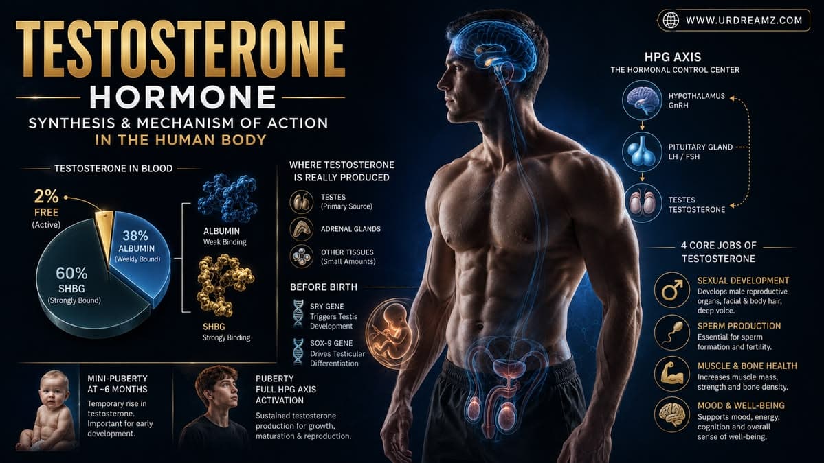
Most people picture testosterone flowing freely and abundantly throughout the bloodstream. The reality is very different: at any given moment, only about 2% of the total testosterone in the body is actually free and biologically active in the blood. The remaining 98% is tightly bound to carrier proteins, held in a kind of reserve rather than circulating freely. If the entire supply of testosterone were somehow released to circulate freely all at once, the resulting hormonal surge would be genuinely destabilizing to the body — this small, tightly regulated free fraction is precisely what allows testosterone to exert its powerful, masculinizing effects on the body in a controlled, sustainable way.
This single fact reframes almost everything most people assume about testosterone. A standard blood test reporting "total testosterone" is measuring both the bound and free portions combined, which is part of why two people can have similar total testosterone readings on paper, yet experience noticeably different symptoms — because what actually matters biologically is how much of that total is genuinely free and available to act on tissue, not simply how much exists in the bloodstream overall.
2. Why the Body Keeps Testosterone So Tightly Controlled
This tight control is not a limitation — it is a deliberate design feature. Testosterone is an extremely potent hormone, and if it were allowed to circulate entirely unbound and unrestricted, its effects on mood, aggression, and physical tissue would be far less predictable and stable. By binding the vast majority of testosterone to carrier proteins and only releasing a small, steady free fraction, the body maintains a much more consistent, well-regulated hormonal environment.
Think of it as similar to how a reservoir works for a city's water supply — rather than letting an entire river flow directly and unpredictably into every household tap, the reservoir holds back the bulk of the supply and releases only a controlled, steady stream as needed. The bound 98% of testosterone functions much like that reservoir, held in reserve and released gradually as carrier proteins release small amounts of testosterone in response to the body's ongoing physiological needs, rather than allowing the hormone's full, potent effect to hit tissue all at once.
3. Male vs Female Bodies: What the Difference Really Comes From
If you compare a typical male body to a typical female body, several consistent differences stand out: male muscles tend to be denser and stronger, while female muscles tend to be comparatively softer; male bones tend to be larger and heavier; males typically carry more muscle mass overall, and even blood volume tends to run higher in males. Behaviorally, males also tend to show more aggression on average, while females tend to show relatively stronger critical thinking tendencies in certain contexts, and risk-taking behavior tends to run higher in males and lower in females.
It is worth being careful with these generalizations — they describe population-level averages and tendencies, not fixed rules that apply to every individual. Substantial overlap exists between male and female populations across virtually every one of these traits, and individual variation within each sex is often larger than the average difference between the sexes. Still, at the population level, these consistent patterns are well-documented across a wide range of studies, and testosterone is understood to be one meaningful contributor among the several factors discussed next.
4. The Five Contributing Factors Behind Male-Female Differences
These differences are not attributable to any single cause. Researchers generally point to a combination of factors: diet, psychological factors, biological factors, and hormonal differences. Testosterone is one significant piece of this broader puzzle — an important contributing factor, but not the sole explanation for every observed difference between male and female bodies and behavior.
5. Myth-Busting: Do Only Males Have Testosterone?
A common misconception holds that testosterone exists only in males and is entirely absent in females. This is incorrect. Females do produce testosterone — simply in significantly smaller quantities compared to males. Testosterone is present and biologically active in both sexes; the difference lies primarily in the quantity produced and circulating, not in its presence or absence.
6. Where Is Testosterone Actually Produced?
Testosterone production in the human body occurs at three distinct anatomical sites, with the relative contribution of each site differing substantially between males and females.
7. Site One: The Testes (About 95%)
In males, the overwhelming majority of testosterone — approximately 95% — is produced in the testes. This makes the testes, by a wide margin, the primary testosterone-producing organ in the male body, and is precisely why conditions or injuries affecting testicular function have such a significant impact on overall testosterone levels.
8. Site Two: The Adrenal Cortex
Both males and females have kidneys, and sitting atop each kidney is a small gland structure containing a region called the adrenal cortex. This region also produces testosterone, contributing a smaller but meaningful portion of the body's total testosterone supply in both sexes.
9. Site Three: The Ovaries
In females, the ovaries also produce testosterone, alongside the contribution from the adrenal cortex described above. Combined, these two sources — the ovaries and the adrenal cortex — represent a female's entire testosterone production, which is why overall testosterone levels in females remain considerably lower than in males, who benefit from the additional, much larger contribution from the testes.
10. Testosterone Is a Steroid Hormone Made From Cholesterol
Testosterone belongs to a category of hormones known as steroid hormones, and its raw building block is cholesterol. This is a genuinely important point: cholesterol, often discussed purely in the negative context of cardiovascular health, is actually an essential raw material the body uses to manufacture testosterone, alongside several other vital steroid hormones. The cholesterol used in this process comes both from what the body produces internally and from dietary intake.
This connection between cholesterol and hormone production is part of why extremely low-cholesterol or low-fat diets, taken to an extreme, can sometimes negatively affect hormone production over time — the body genuinely needs a baseline supply of cholesterol as raw material to keep this hormonal manufacturing process running. This is not an argument for excessive cholesterol intake, which carries its own well-documented cardiovascular risks, but rather a useful reminder that cholesterol is not a purely harmful substance; it serves a genuine, essential biological purpose as the shared starting material for testosterone and several related steroid hormones.
11. The Story Begins Before Birth
To truly understand how testosterone comes to exist in the male body, we need to go all the way back to early fetal development — specifically, to around four to five weeks into pregnancy, when the foundational genetic groundwork for male development is already being laid.
12. XY vs XX: Where Sex Determination Begins
A male fetus develops from a genetic combination involving XY chromosomes, while a female fetus develops from an XX chromosome combination. This chromosomal difference, established at the moment of fertilization, is the genetic starting point from which the entire cascade of male or female physical development eventually unfolds.
13. The SRY Gene: The Master Switch
Present on the Y chromosome of a male fetus is a specific genetic segment called the SRY gene (Sex-determining Region Y). This gene functions as a genuine master switch — its presence and activation is the single genetic trigger that sets male physical development into motion, distinguishing a male fetus's developmental pathway from a female fetus's from this point forward.
The discovery of the SRY gene's specific role was itself a major milestone in reproductive biology, since it pinpointed sex determination down to a single, identifiable genetic segment rather than a vague, broader chromosomal difference. This is also why, in the rare instances where this gene is missing from an otherwise Y-bearing chromosome, or in equally rare cases where it becomes present on an X chromosome, atypical patterns of sexual development can occur — genuine evidence of just how much developmental weight this single gene segment carries.
14. The SOX-9 Gene Takes Over
Once the SRY gene activates, it triggers a second, closely related gene called SOX-9. Because this fetus is genetically male, the SOX-9 gene becomes active as a direct downstream consequence of SRY activation, and it is this SOX-9 activation that actually drives the physical process of male gonadal development from this point forward.
15. Bipotential Gonads: Built to Go Either Way
Before this genetic activation occurs, the developing fetus possesses what are called bipotential gonads — undifferentiated reproductive structures genuinely capable of developing into either male or female anatomy, complete with the early structural precursors, or "canals," for both possible reproductive pathways. Once SOX-9 activates, it specifically halts development of the female reproductive canal pathway and instead drives forward the development of the male testes.
This bipotential starting point is a genuinely elegant feature of early human development — rather than committing irreversibly to one sex or the other from the very earliest stages, the embryo maintains the structural flexibility to develop along either pathway, waiting for the specific genetic signal (or its absence) to determine which set of structures will ultimately be retained and developed, and which will be actively suppressed and regress.
16. By Week 11-12: The Testes Descend Into Place
By around week 11 to 12 of fetal development, this SOX-9-driven process results in the developing testes emerging and becoming structurally established within the fetus. This represents a critical developmental milestone — the physical foundation of the male reproductive system is now genuinely in place, ready for the hormone-producing machinery to begin its work.
17. Meet the Leydig Cells
Within these newly formed fetal testes exist specialized cells called Leydig cells. These cells will become activated shortly after the testes form, and their activation marks the beginning of actual testosterone production within the developing fetus itself — a remarkable process happening well before birth.
18. The Placenta's Role: hCG Activates the Machinery
The trigger that activates these fetal Leydig cells comes from an unexpected source: the mother's own body, specifically the placenta. The placenta releases a hormone called hCG (Human Chorionic Gonadotropin), and once this hCG reaches and activates the Leydig cells within the developing fetus, those cells begin actively producing testosterone from that point forward.
This detail highlights a genuinely fascinating aspect of fetal development: the developing baby is not entirely self-sufficient in producing the hormonal signals needed for its own sexual differentiation — it relies, at least initially, on a hormone supplied through the maternal-placental connection to properly activate this critical developmental process. This same hCG hormone is, notably, the exact substance detected by standard home pregnancy tests, meaning the very hormone confirming a pregnancy is also directly involved in triggering male fetal testosterone production during those crucial early weeks.
19. From Cholesterol to Testosterone Inside the Fetus
Once activated by hCG, the Leydig cells draw upon their internal supply of cholesterol and convert it directly into testosterone. This freshly produced testosterone is then released outward from the Leydig cells, beginning its work of gradually building out the remaining structures of the male reproductive system — including the epididymis, vas deferens, and prostate, each of these ductal structures slowly forming under testosterone's ongoing influence.
20. Building the Male Reproductive Tract
The gradual formation of these internal reproductive structures — the epididymis, vas deferens, and prostate gland — represents testosterone's earliest major construction project within the developing fetus. Each of these structures plays an essential role later in life in the process of sperm maturation, transport, and the production of seminal fluid, and their initial formation during fetal development is entirely dependent on this early testosterone signal.
21. Enter 5-Alpha-Reductase and DHT
At this stage of fetal development, testosterone itself is acted upon by a specific enzyme called 5-alpha-reductase. This enzyme converts a portion of the available testosterone into an even more potent hormone called DHT (Dihydrotestosterone). DHT is significantly more powerful than testosterone itself in certain tissues, and it is also, notably, one of the primary hormones responsible for male pattern hair loss later in adult life.
The reason DHT is considered more potent than testosterone in specific tissues comes down to how strongly each hormone binds to the same underlying receptor. DHT binds to androgen receptors with significantly greater affinity and produces a stronger, longer-lasting activation of those receptors compared to testosterone itself, even though both hormones work through fundamentally the same receptor system. This is precisely why certain tissues — including hair follicles and external genital tissue — respond so much more strongly to DHT specifically, rather than to the broader pool of circulating testosterone.
22. DHT Finishes the Job: External Genitalia and the Scrotum
Once activated within the developing fetus, DHT takes over responsibility for forming the external reproductive organs, along with the scrotum — the external sac that will house the testes. Essentially, while testosterone builds the internal ductal structures, DHT completes the external anatomical structure of the male reproductive system, finishing what testosterone started.
23. Six Months After Birth: Mini-Puberty
Once the baby is born, a curious and often-overlooked event occurs approximately six months later: a phenomenon called mini-puberty begins in the infant's brain. This can be understood as a small-scale, temporary preview of full puberty — a partial activation of the same hormonal system that will later drive full adolescent puberty years down the line.
Mini-puberty occurs in both male and female infants, though its specific hormonal profile and physical effects differ somewhat between the sexes. In male infants specifically, this period is understood to play a meaningful role in the further maturation and calibration of testicular tissue, including the ongoing development of the very Leydig and Sertoli cell populations that will become critical again years later, during full puberty. Researchers continue to study this window closely, since it appears to offer a rare, naturally occurring opportunity to observe key reproductive hormone activity in infancy, well before the more dramatic and sustained hormonal changes of adolescence begin.
24. Why Mini-Puberty Matters
During this period, the infant's hypothalamus receives an internal signal to release GnRH (Gonadotropin-Releasing Hormone). Just below the hypothalamus sits the pituitary gland, which becomes activated by this GnRH signal and, in turn, releases two important hormones: LH (Luteinizing Hormone) and FSH (Follicle-Stimulating Hormone). These hormones travel down to the developing reproductive "factory," helping ensure it develops properly and completely — a process understood to matter significantly for future reproductive function later in life.
25. The Long Pause Before Real Puberty
After this six-month mini-puberty window passes, this entire hormonal system goes quiet — entering a lengthy pause that lasts for roughly twelve to thirteen years. During this extended period of childhood, the GnRH-LH-FSH signaling system remains largely dormant, only reactivating much later, at the actual onset of puberty.
26. Age 13-14: The System Restarts
Once a child reaches approximately 13 to 14 years of age, this same hormonal signaling system reactivates — the hypothalamus begins releasing GnRH once again, restarting the exact same cascade that had briefly appeared during infancy's mini-puberty, only now on a much larger, sustained scale that will drive the child through full adolescent puberty.
27. Why GnRH Must Pulse, Not Flow Continuously
Critically, GnRH release during puberty does not occur as a constant, continuous stream — it is released in distinct pulses, occurring roughly every one to one-and-a-half to two hours. This pulsatile release pattern is essential; if GnRH were instead released continuously and constantly rather than in these carefully spaced pulses, this would actually lead to reproductive dysfunction and infertility later in life, since the downstream pituitary receptors depend specifically on this rhythmic, intermittent signaling pattern to function correctly.
This is actually the exact biological principle exploited by certain medical treatments — continuous, rather than pulsatile, GnRH-based medications are deliberately used in some clinical settings specifically to suppress reproductive hormone production, precisely because sustained, non-pulsatile GnRH exposure causes the pituitary's GnRH receptors to become desensitized and effectively shut down, rather than stimulated. This same mechanism, when it occurs naturally rather than through medication, is exactly why disrupted or absent GnRH pulsing during puberty can result in delayed puberty or reduced fertility.
28. LH and FSH Travel to the Factory
Each of these pulsatile GnRH signals prompts the pituitary gland to release LH and FSH into the bloodstream, where these hormones travel down to the male reproductive "factory" — the testes — to carry out their specific, distinct jobs.
29. Meet the Sertoli Cells: Nurse Cells for Sperm
Upon reaching the testes, FSH specifically binds to and activates Sertoli cells. These Sertoli cells function as dedicated "nurse cells," directly nourishing and supporting the developing precursor sperm cells — specifically the spermatid and spermatocyte cells, which are the earlier developmental stages that will eventually mature into fully functional sperm. Without adequate Sertoli cell support, these developing sperm precursors cannot properly mature.
Sertoli cells perform several additional supporting roles beyond simple nourishment. They form tight physical junctions with one another that create what is known as the blood-testis barrier, a protective structure that shields developing sperm cells from the immune system, which would otherwise potentially recognize these genetically unique cells as foreign, given that sperm cells only carry half the normal genetic material and continue developing well after the immune system has already established its sense of "self" earlier in life. This immune-shielding function, alongside their nourishing role, makes Sertoli cells absolutely essential to successful, ongoing sperm production throughout adult male life.
30. Meet the Leydig Cells Again: The Testosterone Factory
LH, meanwhile, travels to and binds specifically to the outer boundary cells of the testes — the same Leydig cells introduced earlier in the fetal development section. Upon binding, LH converts these Leydig cells into active testosterone-producing units once again, and it is this LH-driven activation of Leydig cells during puberty that produces the surge of testosterone responsible for the wide range of physical changes associated with male adolescence.
It's worth appreciating the symmetry here: the exact same LH-Leydig cell relationship that first produced testosterone weeks into fetal development is reactivated, years later, to drive the far more visible and dramatic transformation of puberty — deepening of the voice, growth of facial and body hair, increased height and muscle mass, and the full maturation of the reproductive system. The underlying cellular machinery essentially remains the same throughout a male's life; what changes is simply how strongly and consistently that machinery gets switched on.
31. Still Only 2% Free — Even After Puberty
Even after puberty is complete and the body's testosterone-producing machinery is fully operational, the same fundamental principle discussed at the beginning of this guide still holds true: testosterone does not simply circulate freely throughout the bloodstream in its entirety. Only about 2% remains free and biologically active — if the entire supply were to circulate freely and unrestricted, it would actually result in infertility, since the body's reproductive system depends on this carefully regulated balance rather than an unrestrained flood of the hormone.
32. SHBG and Albumin: The Hormone's Two Carriers
Once testosterone is released from the Leydig cells into the bloodstream, it does not travel alone — the vast majority of it becomes bound to two specific carrier proteins: SHBG (Sex Hormone-Binding Globulin) and Albumin.
| Carrier / State | Approximate Share of Total Testosterone |
|---|---|
| Bound to SHBG | ~60% |
| Bound to Albumin | ~38% |
| Free (unbound) testosterone | ~2% |
33. Why This Binding System Exists
SHBG holds onto approximately 60% of total testosterone, while Albumin holds onto roughly 38%, leaving only the remaining 2% freely circulating and biologically active in the blood at any given moment. This remaining free 2% is precisely the portion responsible for driving sperm production, libido (sexual desire), muscle and bone health, and increased blood cell production — every one of testosterone's major physiological effects traces back to this small, carefully regulated free fraction.
It is worth noting that these two carrier proteins behave somewhat differently in how tightly they bind their testosterone cargo. SHBG binds testosterone very tightly, making that bound portion essentially unavailable for immediate biological action. Albumin, on the other hand, binds testosterone somewhat more loosely, meaning albumin-bound testosterone can, under certain conditions, become available to tissues relatively quickly. Because of this distinction, some clinicians and researchers refer to the combination of free testosterone plus albumin-bound testosterone together as "bioavailable testosterone," recognizing that this broader category represents the portion of the body's total testosterone supply that is realistically available for use, distinct from the more tightly locked-away SHBG-bound portion.
34. DHT's Second Act: Facial and Body Hair
Beyond its role during fetal development, the free testosterone circulating after puberty continues to be converted into DHT throughout adult life. Once puberty is reached, this DHT becomes specifically responsible for the growth of facial hair, such as a beard and mustache, along with broader body hair growth patterns associated with male adolescence and adulthood.
This same DHT pathway, later in adult life, is also directly implicated in male pattern hair loss, sometimes called androgenic alopecia. In genetically predisposed hair follicles, particularly at the hairline and crown of the scalp, DHT gradually causes a process called follicular miniaturization, in which hair follicles shrink over successive growth cycles, eventually producing progressively finer, shorter hairs before follicles stop producing visible hair altogether. Interestingly, this is the same DHT responsible for facial and body hair growth elsewhere on the body, illustrating how the very same hormone can produce dramatically different, even opposite, effects depending entirely on which specific tissue and follicle type it is acting upon.
35. Testosterone, the Kidneys, and EPO
When free testosterone circulating in the blood eventually reaches the kidneys, it stimulates the production of another important hormone called EPO (Erythropoietin). EPO's specific job is to travel into the bone marrow, where it accelerates and speeds up the production of new red blood cells — a process called erythropoiesis.
36. Why Men Have Higher Blood Counts Than Women
This testosterone-driven EPO stimulation is precisely why male blood hemoglobin levels typically run higher than female levels — commonly cited as approximately 15 to 17 g/dL in males, compared to approximately 12 to 14 g/dL in females. This consistent difference in normal reference ranges between the sexes traces directly back to testosterone's stimulating effect on red blood cell production via the kidney-EPO-bone marrow pathway.
37. The Four Core Functions of Testosterone
Bringing everything together, the free testosterone released from the Leydig cells ultimately performs four essential, defining functions throughout the body:
- Protein Synthesis: Testosterone directly supports the body's ability to build and repair protein-based tissue, playing a central role in muscle growth and overall tissue maintenance.
- Bone Health: Testosterone contributes to maintaining bone density and strength, helping protect against bone thinning and fragility over time.
- Erythropoiesis: Through its stimulation of EPO release from the kidneys, testosterone drives increased red blood cell production in the bone marrow, contributing to the higher blood counts typically observed in males.
- Spermatogenesis: Working alongside FSH and the Sertoli cells, testosterone is essential for the ongoing production and maturation of sperm cells within the testes.
38. Testosterone Production and Regulation: Summary Table
| Stage / System | Key Players | Outcome |
|---|---|---|
| Fetal Development (Week 4-12) | SRY gene, SOX-9 gene, Leydig cells, hCG | Testes form; internal reproductive ducts built |
| Fetal DHT Formation | 5-alpha-reductase | External genitalia and scrotum formed |
| Mini-Puberty (6 months old) | GnRH, LH, FSH | Reproductive system further matured |
| Puberty (13-14 years) | Pulsatile GnRH, LH, FSH, Sertoli cells, Leydig cells | Full testosterone production begins; adolescent changes |
| Adult Circulation | SHBG (60%), Albumin (38%), Free testosterone (2%) | Protein synthesis, bone health, erythropoiesis, spermatogenesis |
39. Frequently Asked Questions
- Q1: How much testosterone actually circulates freely in the blood?
- Only about 2% of total testosterone circulates freely and is biologically active; the remaining 98% is bound to carrier proteins SHBG and Albumin.
- Q2: Do females produce testosterone too?
- Yes. Females produce testosterone from the ovaries and adrenal cortex, though in significantly smaller amounts than males.
- Q3: Where is most testosterone produced in males?
- Approximately 95% of testosterone in males is produced in the testes, specifically by Leydig cells.
- Q4: What raw material is testosterone made from?
- Testosterone is a steroid hormone synthesized from cholesterol.
- Q5: What is the SRY gene?
- The SRY gene is a segment on the Y chromosome that acts as the master genetic switch triggering male development in a fetus.
- Q6: What is the role of the SOX-9 gene?
- SOX-9 is activated following SRY activation and drives the physical development of the testes from bipotential gonadal tissue.
- Q7: What triggers testosterone production in a developing fetus?
- The placenta releases hCG (Human Chorionic Gonadotropin), which activates fetal Leydig cells to begin producing testosterone.
- Q8: What is DHT and why does it matter?
- DHT (Dihydrotestosterone) is a more potent hormone converted from testosterone via the enzyme 5-alpha-reductase; it forms external genitalia in the fetus and is later responsible for facial hair growth and male pattern hair loss.
- Q9: What is mini-puberty?
- Mini-puberty is a brief hormonal activation occurring around six months after birth, involving GnRH, LH, and FSH, which helps further develop the reproductive system before the system goes dormant until actual puberty.
- Q10: Why is GnRH released in pulses rather than continuously?
- Pulsatile GnRH release is required for proper pituitary gland response; continuous, non-pulsatile release would actually lead to reduced fertility rather than supporting it.
- Q11: What do Sertoli cells do?
- Sertoli cells, activated by FSH, act as nurse cells that nourish and support developing sperm precursor cells (spermatids and spermatocytes) as they mature.
- Q12: What do Leydig cells do?
- Leydig cells, activated by LH, are responsible for producing testosterone from cholesterol, both during fetal development and throughout adult life.
- Q13: What are SHBG and Albumin?
- SHBG and Albumin are carrier proteins that bind approximately 60% and 38% of circulating testosterone respectively, leaving only about 2% free and active.
- Q14: Why do men typically have higher blood counts than women?
- Free testosterone stimulates the kidneys to release EPO (Erythropoietin), which accelerates red blood cell production in the bone marrow, resulting in higher typical hemoglobin levels in men.
- Q15: What are the four main functions of testosterone?
- Testosterone's four core functions are protein synthesis, maintaining bone health, stimulating erythropoiesis (red blood cell production), and supporting spermatogenesis (sperm production).
40. Conclusion
Testosterone's story begins remarkably early — weeks into fetal development, when a single genetic switch on the Y chromosome sets off a cascade involving the SOX-9 gene, the formation of the Leydig cells, and a hormonal assist from the mother's own placenta, ultimately building both the internal and external structures of the male reproductive system before birth. That same hormonal system briefly reactivates at six months of age in a mini-puberty, goes dormant for over a decade, and then fully reawakens at adolescence through a precisely pulsatile signal from the brain. Throughout adult life, this hormone remains tightly regulated, with 98% held in reserve by carrier proteins and only a small 2% free fraction responsible for everything from muscle and bone strength to red blood cell production and fertility itself. Understanding this full arc — from a single gene before birth to the four essential functions testosterone performs in adulthood — reveals just how precisely engineered, and how far from a simple "free-flowing" hormone, testosterone actually is.