Sperm and Ovum Fusion: The Exact Molecular Process of Fertilization
In our previous deep dive, we traced the sperm cell itself — every structural component, from its enzyme-loaded acrosome to its mitochondria-packed midpiece — and followed it right up to the very threshold of the egg. This article picks up exactly where that one left off, and goes even deeper: into the precise molecular choreography that unfolds the instant sperm and egg finally touch. What happens in the seconds, minutes, and hours after that first contact is one of the most tightly regulated, exquisitely timed sequences of events in all of human biology — a cascade involving specific enzymes, a single sperm-delivered protein that triggers a rhythmic wave of calcium through the entire egg, an emergency defense system that locks the door behind the winning sperm within moments, and the formation of two entirely separate genetic packages that must find each other and combine before a single new cell — a zygote — can begin to divide.
This is a research-based, medically accurate, step-by-step breakdown of that exact process: sperm penetration, the acrosome reaction, calcium signaling, cortical granule release, the prevention of polyspermy, pronuclear formation, and the very first cleavage. We'll also look at how embryologists in fertility clinics around the world use this exact molecular sequence, in practice, every single day, to confirm whether fertilization in an IVF laboratory has genuinely succeeded.
Quick Answer
Last updated: September 2026
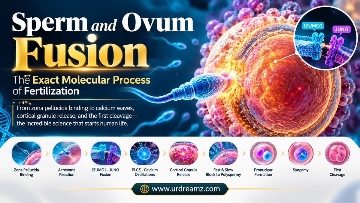
Sperm-ovum fusion (fertilization) begins when a capacitated sperm binds to and penetrates the egg's outer layers — the cumulus oophorus, corona radiata, and zona pellucida — aided by the acrosome reaction, which releases the enzymes hyaluronidase and acrosin. The sperm's IZUMO1 protein then binds to the egg's JUNO receptor, triggering membrane fusion. At the moment of fusion, the sperm delivers a specific enzyme called PLCζ (PLC-zeta) into the egg's cytoplasm, which triggers a release of calcium from internal stores, producing a repeating pattern known as calcium oscillations. This calcium signal does several things simultaneously: it silences the egg's Cytostatic Factor (CSF), which had held it arrested at Metaphase II, allowing meiosis to finish and the second polar body to be released; it triggers cortical granules to release enzymes that harden the zona pellucida (the "slow block to polyspermy"), working alongside a rapid membrane depolarization (the "fast block"), preventing any additional sperm from entering. The sperm and egg genetic material then each form a separate pronucleus; the sperm's DNA decondenses from its tightly packed protamine state, and both pronuclei come together in a process called syngamy, initiating the zygote's first mitotic division, or cleavage.
1. From Two Cells to One: What "Fertilization" Actually Means Molecularly
At its simplest definition, fertilization is the union of a sperm and an egg to form a single new cell called a zygote. But that one-sentence definition hides an extraordinarily intricate molecular process — a tightly sequenced, multi-stage cascade that must execute correctly, in exact order, for a viable, chromosomally normal zygote to form at all. This article breaks that entire cascade down, stage by stage, exactly as it unfolds at the cellular and molecular level, from the moment a sperm first makes contact with the outermost layer of cells surrounding the egg, all the way through to the zygote's first cell division.
2. Picking Up Where We Left Off: A Quick Bridge From Sperm Anatomy
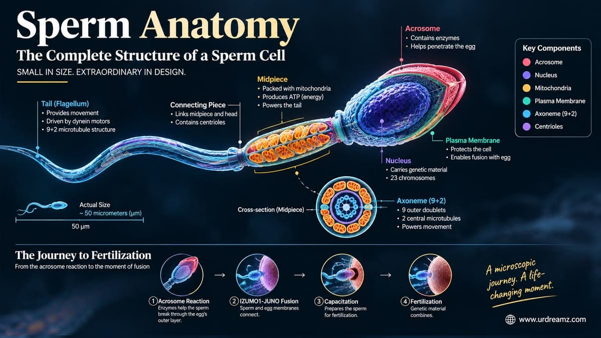
Our earlier article on sperm anatomy covered the sperm cell's complete structure in detail — its acrosome-loaded head, its mitochondria-packed midpiece, and the axoneme-driven tail that propels it forward, along with the process of capacitation that primes it for this exact moment. This article assumes that background and picks up the story at the precise point that article left open: a fully capacitated, hyperactivated sperm cell has now arrived in the vicinity of a secondary oocyte within the ampulla of the fallopian tube, ready to begin the final, decisive stretch of its journey.
3. The Cumulus Oophorus: The Outer Cloud of Cells Around the Egg
Before a sperm ever reaches the egg's actual protective shell, it must first pass through a loose, cloud-like cluster of follicular cells called the cumulus oophorus. This structure is released from the ovary alongside the egg at ovulation and surrounds it throughout its journey down the fallopian tube. The cumulus oophorus is held together by a matrix rich in hyaluronic acid, and its outermost boundary blends into a more tightly packed inner layer of cells, called the corona radiata, sitting directly against the egg's next protective barrier. Sperm must navigate through this looser outer cloud first, using both their swimming motion and, as we'll cover shortly, enzymatic assistance, before ever reaching the more tightly organized layers beneath it.
3b. Where the Egg Was Before All of This: A Brief Note on Ovulation
It's worth briefly grounding this entire process in what happens just before the sperm ever arrives. Around the midpoint of a typical menstrual cycle, a surge in luteinizing hormone triggers ovulation, the release of a secondary oocyte from a mature ovarian follicle. At the point of release, this egg is not yet fully mature in the sense this article requires — it is arrested partway through its second meiotic division, a paused state we'll return to in detail later in this article, and it carries with it the cumulus oophorus and corona radiata layers described above, having formed a close relationship with these surrounding cells during its extended development within the ovary. This released egg is then swept into the fallopian tube by finger-like projections called fimbriae, beginning its own journey toward the ampulla, the specific region where, as covered later in this article, fertilization actually takes place.
4. A Quick Refresher: Corona Radiata and Zona Pellucida
| Layer | Position | Composition | Broken Down By |
|---|---|---|---|
| Cumulus Oophorus | Outermost, loosely organized layer | Follicular cells in a hyaluronic acid matrix | Sperm motility and hyaluronidase activity |
| Corona Radiata | Inner portion of the cumulus complex, directly adjacent to the zona pellucida | More tightly packed follicular cells | Hyaluronidase (released during the acrosome reaction) |
| Zona Pellucida | Innermost, directly surrounding the egg's own membrane | A structural layer built from specific zona pellucida glycoproteins | Acrosin and continued sperm motility |
5. First Contact: Sperm Binding to the Zona Pellucida
Once a sperm has made its way through the cumulus and corona layers, it makes direct contact with the zona pellucida — and this contact is not random or generic. The zona pellucida is built from a specific set of glycoproteins, and sperm-surface receptor molecules recognize and bind to these zona glycoproteins in a manner that displays meaningful species specificity, helping ensure that, under normal biological conditions, fertilization occurs between compatible gametes. This binding step is what triggers the next critical event in the sequence: the acrosome reaction.
5b. How Sperm "Recognize" the Right Egg: Species Specificity
It's worth understanding why zona pellucida binding matters so much beyond simply being a mechanical first step. The zona pellucida's glycoprotein structure differs meaningfully between species, and sperm-surface receptor proteins have evolved to recognize the specific glycoprotein patterns of their own species' zona pellucida. This built-in molecular specificity is a significant part of what normally prevents cross-species fertilization under natural conditions — a sperm cell from one species generally cannot successfully bind to and penetrate the zona pellucida of an egg from an unrelated species, because the specific molecular "fit" simply isn't there. Researchers continue to refine exactly which zona proteins and sperm receptors are most responsible for this specificity, since earlier models proposing a single dominant receptor-ligand pair have been revised as more detailed molecular studies have emerged, but the overall principle — that this binding step carries genuine biological specificity, not just mechanical proximity — remains well established.
6. The Acrosome Reaction, Revisited in Full Molecular Detail
As covered in our earlier sperm anatomy article, the acrosome reaction is the process by which the sperm's acrosomal cap releases its stored enzymes. Here's the fuller molecular picture: contact with the zona pellucida triggers a calcium influx into the sperm cell itself, which in turn causes the outer acrosomal membrane to fuse with the sperm's overlying plasma membrane at multiple points. This fusion creates openings through which the acrosome's contents — principally hyaluronidase and acrosin — are released directly onto the egg's protective layers. This reaction is irreversible and single-use: once triggered, a sperm cell cannot "reload" its acrosome, meaning the timing of this reaction, occurring at exactly the right proximity to the egg, is genuinely critical to a successful outcome.
Contact
Into Sperm
Fuses With Plasma Membrane
(Hyaluronidase, Acrosin)
7. Tunneling Through: Mechanical and Enzymatic Zona Penetration
Penetrating the zona pellucida isn't purely a chemical process — it's a combination of enzymatic digestion from acrosin and the sperm's own continued, hyperactivated swimming motion, discussed in our previous article, physically driving it forward through the partially dissolved barrier. This combined mechanical-and-enzymatic approach carves a narrow tunnel through the zona pellucida's structural mesh, and the sperm advances through this tunnel until it finally reaches the innermost boundary of the zona, immediately adjacent to the egg's own outer membrane.
8. The Perivitelline Space: The Final Gap Before Fusion
Once through the zona pellucida, the sperm enters a narrow, fluid-filled gap called the perivitelline space, sitting directly between the inner surface of the zona pellucida and the egg's own plasma membrane (also called the oolemma). This is the final physical gap the sperm must cross, and it's within this space that the sperm's head makes its first direct contact with the egg's actual cell membrane — the setting for the fusion event covered in the next section.
9. IZUMO1 Meets JUNO: The Fusion Handshake, in Detail
As introduced in our sperm anatomy article, successful membrane fusion depends on a specific molecular interaction between IZUMO1, a protein on the sperm's surface, and JUNO, its corresponding receptor on the egg's surface. Research has established that this interaction isn't simply about physical adhesion — it functions as a genuinely essential recognition and activation step, without which membrane fusion cannot proceed, regardless of how successfully the sperm navigated every prior stage of the journey. Interestingly, research has also found that shortly after the first successful fusion event, JUNO is rapidly removed from the egg's surface — a mechanism that appears to contribute directly to the block to polyspermy discussed later in this article, closing this specific "lock" almost as quickly as it was opened.
10. The Moment of Fusion: Sperm Content Enters the Egg
Once IZUMO1 and JUNO bind, the plasma membranes of the sperm and egg fuse into a single, continuous membrane. This is the genuine cellular threshold of fertilization: the sperm's entire content — its nucleus, its midpiece with its mitochondria, and critically, a specific enzyme it has been carrying since maturation — now enters directly into the egg's cytoplasm (also referred to as the ooplasm). It's worth noting that while the sperm's mitochondria do enter the egg, they are typically actively targeted for degradation shortly afterward, which is part of why mitochondrial DNA is inherited almost exclusively from the mother rather than the father — a separate, fascinating detail beyond the scope of this article's central focus.
10b. Why You Only Inherit Mitochondria From Your Mother
The brief mention of sperm mitochondrial degradation in the previous section deserves a slightly fuller explanation, since it's a genuinely well-known and important fact in human genetics. Although the sperm's midpiece, packed with mitochondria as covered in our earlier sperm anatomy article, does physically enter the egg at fusion, these paternal mitochondria are specifically tagged for destruction shortly afterward, through a regulated cellular process involving ubiquitin-mediated degradation, and are eliminated well before the embryo develops significantly further. As a result, essentially all of the mitochondria present in a developing embryo — and therefore in the resulting person, throughout their entire life — originate from the egg, meaning mitochondrial DNA is passed down along the maternal line only. This specific inheritance pattern is precisely why mitochondrial DNA analysis has become such a valuable tool in genetics and ancestry research: it provides a clean, uninterrupted maternal lineage record, entirely unaffected by paternal contribution at each generation.
11. PLCζ: The Molecule the Sperm Smuggles In
Among everything the sperm delivers into the egg at the moment of fusion, one molecule stands out as the true trigger for everything that follows: a sperm-specific enzyme called PLCζ (PLC-zeta, or phospholipase C zeta). This enzyme is not present in the egg itself — it's a genuinely sperm-exclusive protein, carried into the egg specifically at the moment of fusion, and its arrival sets off the entire downstream signaling cascade responsible for "activating" the egg out of its long-held dormant state.
12. IP3 and the Calcium Store: How PLCζ Triggers the Wave
Once inside the egg's cytoplasm, PLCζ acts on a membrane lipid called PIP2, breaking it down into a smaller signaling molecule called inositol trisphosphate, or IP3. IP3 then travels to and binds receptors on the egg's endoplasmic reticulum — an internal membrane network that, in the egg, functions as a substantial internal storage depot for calcium ions. This binding causes the endoplasmic reticulum to release a significant pulse of calcium directly into the egg's cytoplasm, marking the true beginning of the calcium signal that drives every subsequent stage of egg activation covered in this article.
PLCζ
PIP2 Into IP3
Receptors
From Storage
13. Calcium Oscillations: Not One Spike, But a Rhythm
One of the most striking and well-documented features of egg activation in mammals is that this calcium release doesn't happen as a single, one-time spike — it occurs as a repeating series of rhythmic pulses called calcium oscillations, continuing for a period of several hours after fusion. Each individual pulse follows a similar pattern: calcium levels rise sharply, then gradually return to baseline, before rising again in the next wave. This oscillatory pattern, rather than a single flat signal, is now understood to be biologically essential — sustained, rhythmic calcium signaling activates specific downstream enzymes more effectively and reliably than a single transient spike would, and research has shown that disrupted or abnormal oscillation patterns are associated with fertilization and early embryo development failures, making this rhythmic quality a genuinely critical feature rather than an incidental biological detail.
*Simplified illustrative representation of the repeating calcium oscillation pattern; actual patterns vary and are measured via specialized calcium-imaging techniques.
14. What Calcium Actually Switches On Inside the Egg
This calcium signal isn't a vague, generic "activation" — it triggers several specific, well-defined downstream processes simultaneously, each of which this article covers individually in the sections ahead. Broadly, the rising calcium levels activate calcium/calmodulin-dependent protein kinase II (CaMKII), an enzyme that in turn sets off a chain of events including the release of cortical granules, the resumption of the cell cycle that had been paused for years within the egg, and the metabolic changes needed to support the earliest stages of embryonic development. In effect, this single molecular signal — the calcium wave triggered by the sperm's delivery of PLCζ — functions as the master switch that transforms a dormant, arrested egg cell into an actively developing embryo.
15. CSF and Metaphase Arrest: Why the Egg Was "Paused" All Along
To understand why calcium's effect is so significant, it helps to understand the state the egg was actually in just before fertilization. Unlike most cells, a mature human egg is held in a genuinely paused, arrested state at a specific point in its own cell division process, called Metaphase II of meiosis — essentially frozen partway through completing its final division. This pause is maintained by a group of molecules collectively referred to as Cytostatic Factor, or CSF, a signaling pathway that actively prevents the cell cycle from proceeding any further until an appropriate trigger arrives. Without fertilization, the egg would simply remain arrested at this exact stage indefinitely, eventually degrading without ever completing its own developmental process.
16. Breaking the Pause: How Calcium Silences CSF
The calcium oscillations triggered by PLCζ are precisely what breaks this long-held pause. Through the CaMKII pathway mentioned earlier, rising calcium levels ultimately lead to the activation of a protein complex called the Anaphase-Promoting Complex, which in turn targets specific regulatory proteins within the CSF pathway for degradation. With CSF effectively silenced, the cell cycle "brake" is released, and the egg is finally free to complete the meiotic division it had been paused partway through, sometimes for years, depending on how long that particular egg had been waiting since ovulation was first triggered during the woman's monthly cycle.
17. Cortical Granules: The Egg's Emergency Response Team
Simultaneously with silencing CSF, the same calcium signal triggers another critical, time-sensitive process: the release of cortical granules. These are small, enzyme-filled vesicles positioned just beneath the egg's outer membrane, arranged around its entire circumference, essentially standing ready for exactly this moment. Upon the calcium trigger, these granules undergo exocytosis — fusing with the egg's outer membrane and releasing their enzymatic contents into the perivitelline space and onto the zona pellucida, in a process called the cortical reaction. This release happens remarkably quickly, within seconds of the initial calcium rise, reflecting just how time-critical this specific defensive response actually is.
18. Zona Hardening: The Slow Block to Polyspermy
The enzymes released from cortical granules chemically modify the structure of the zona pellucida's glycoproteins, most notably cleaving a specific zona protein in a way that alters its physical properties, making the entire zona pellucida structure significantly tougher, less permeable, and effectively resistant to further sperm penetration. This process, often called zona hardening, takes a short but meaningful amount of time to complete fully, which is why it's frequently referred to as the "slow block" to polyspermy — slower than the near-instantaneous electrical block described next, but no less important as a durable, lasting barrier against any additional sperm that might still be attempting to penetrate the zona in the following minutes.
19. The Fast Block: Membrane Depolarization
Working alongside the slower, zona-based defense is a much faster, though shorter-lived, electrical mechanism. The moment the first sperm fuses with the egg's membrane, ion channels open, briefly changing the electrical charge across the egg's plasma membrane — a rapid depolarization that, for a short window immediately following fusion, makes the membrane itself less receptive to additional sperm fusion events. This fast electrical block acts as an immediate, first-line defense, buying critical time for the slower, more durable zona-hardening process described in the previous section to complete and take over as the primary, lasting barrier.
Fuses
(Membrane Depolarization)
Triggers Cortical Reaction
(Zona Hardening)
20. When the System Fails: Polyspermy and Triploidy
Despite these layered defenses, the block to polyspermy occasionally fails, allowing more than one sperm to fertilize the same egg — a rare event called polyspermy, or dispermy when specifically two sperm are involved. When this happens, the resulting embryo ends up with three complete sets of chromosomes instead of the normal two, a condition called triploidy, since it received one set from the egg and two separate sets from two different sperm. This extra genetic material severely disrupts normal cell division, and the overwhelming majority of triploid embryos fail to develop and result in very early pregnancy loss. In extraordinarily rare, well-documented cases, a small number of surviving pregnancies from this kind of unusual fertilization event have resulted in what researchers call sesquizygotic, or "semi-identical," twins — a genuinely fascinating and rare finding that has been reported in international medical literature, illustrating just how tightly regulated and evolutionarily important the normal block-to-polyspermy mechanism really is.
20b. Why Fertilization Creates Genetic Diversity, Not Just Genetic Combination
Beyond simply combining two sets of chromosomes, the process covered throughout this article is also the reason every human being, aside from identical twins, is genetically unique. During the meiotic divisions that produce both sperm and egg, a process called genetic recombination shuffles segments of chromosomes between maternal and paternal copies before they're separated into individual gametes, meaning no two sperm cells from the same man, and no two eggs from the same woman, carry an identical genetic combination. Combined with the essentially random nature of exactly which sperm, among the hundreds of millions released, ultimately succeeds in completing the entire sequence described in this article — zona binding, the acrosome reaction, membrane fusion, and successful calcium-triggered activation — the resulting zygote represents one specific, unrepeatable genetic combination out of an astronomically large number of possible outcomes. This is part of why the tightly regulated, single-winner design of the fertilization process covered throughout this article matters so much biologically: it's not simply about combining DNA, but about ensuring that combination happens through a single, cleanly executed event, producing one genetically coherent, viable new individual rather than a chaotic genetic mixture.
21. Completing Meiosis: The Second Polar Body
With CSF silenced and the metaphase arrest lifted, the egg is finally free to complete the second meiotic division it had been paused within. This division is highly asymmetric: rather than splitting into two similarly sized cells, the egg retains almost all of its cytoplasm and nutrient reserves for itself, while extruding a small, genetically complete but cytoplasm-poor cell called the second polar body, which contains a duplicate set of chromosomes but is not capable of further development. This asymmetric division ensures the vast majority of the egg's valuable cytoplasmic resources remain available to support the developing embryo, rather than being evenly split between two cells the way an ordinary cell division typically would be.
22. Pronuclear Formation: Two Genomes, Two Separate Envelopes
Following the completion of meiosis, the egg's own remaining chromosomes become enclosed within a newly formed nuclear envelope, creating what's called the female pronucleus. Meanwhile, on the other side of the now-fertilized egg, the sperm's genetic material undergoes its own parallel transformation, forming the male pronucleus, described in more detail in the next section. Importantly, these two pronuclei remain entirely separate structures at this stage — they do not immediately merge into one — each visibly distinct under a microscope, a detail that turns out to have significant practical, clinical relevance, covered later in this article.
22b. Tracking the Chromosome Count Through Every Stage
Given how much this article has referenced chromosome numbers throughout, it helps to see the complete picture laid out clearly in one place, tracking the transition from haploid (n) to diploid (2n) at each key stage of the process.
| Stage | Chromosome Set | Notes |
|---|---|---|
| Mature Sperm | 23 chromosomes (n, haploid) | Half the normal chromosome number, ready to combine with the egg's set |
| Egg Before Fertilization (Metaphase II arrest) | 23 chromosomes (n, haploid), duplicated | Paused mid-division, awaiting the fertilization trigger |
| Second Polar Body | 23 chromosomes (n, haploid) | Discarded during asymmetric meiotic division; not involved in development |
| Female Pronucleus | 23 chromosomes (n, haploid) | The egg's own completed genetic contribution |
| Male Pronucleus | 23 chromosomes (n, haploid) | The sperm's decondensed genetic contribution |
| Zygote (After Syngamy) | 46 chromosomes (2n, diploid) | The complete, combined genetic blueprint of a new individual |
This table captures, in a single glance, precisely why every stage covered in this article matters: each step exists specifically to ensure that two separate haploid sets, one from each parent, are correctly and safely combined into exactly one complete diploid set, no more and no less, forming the genetic foundation of a new, viable zygote.
23. Sperm Decondensation: From Protamines Back to Histones
Recall from our sperm anatomy article that the sperm's DNA is packaged extremely tightly using proteins called protamines, reinforced by disulfide bridges, specifically to survive its long journey intact. Once inside the egg, this tightly compacted state must be actively reversed. Enzymes and factors within the egg's cytoplasm break the disulfide bonds holding the protamine-packed DNA together, and the protamines are gradually replaced with maternally supplied histones — the same class of DNA-packaging protein used by essentially every other cell in the body. As this exchange occurs, the sperm's nucleus visibly swells and decondenses, transforming from its tightly compacted, transport-ready state into a properly organized, functional nucleus capable of participating in the developing embryo's genetic activity — the male pronucleus referenced in the previous section.
24. Centrioles and the Sperm Aster: Guiding the Pronuclei Together
As covered in our sperm anatomy article, the sperm contributes something the egg itself typically lacks in a fully functional state: a centriole. Shortly after fertilization, this sperm-derived centriole organizes a radiating network of microtubules called the sperm aster, extending outward through the egg's cytoplasm. This structure plays a genuinely critical mechanical role: it physically helps draw the male and female pronuclei toward each other from their initially separate positions within the fertilized egg, guiding them into close proximity in preparation for the next stage of this process.
25. Syngamy: The First Mitotic Division Begins
Once the male and female pronuclei have been drawn together by the sperm aster, their nuclear envelopes break down, and the two sets of chromosomes — 23 from the mother, 23 from the father — align together on a shared mitotic spindle in preparation for the zygote's very first cell division. This overall process, through which the genetic material from both parents is brought together to function as a single, unified genome, is referred to as syngamy. It's worth noting a genuinely interesting technical detail: the two pronuclei don't necessarily fuse together into a single nucleus first the way some textbook diagrams simplify it — in humans, their chromosomes typically come together directly on the first mitotic spindle, with the full, unified diploid nucleus only properly forming after this first division is complete.
26. The Zygote and First Cleavage
The single cell formed once sperm and egg genetic material have combined is called a zygote — genetically complete, carrying a full, unique 46-chromosome combination distinct from either parent alone. Roughly 24 to 30 hours after fertilization, this zygote undergoes its first mitotic division, called cleavage, splitting into two cells. This marks the true beginning of embryonic development, and it's followed by a rapid, repeating sequence of further divisions over the following days as the embryo continues developing on its journey toward the uterus.
| Approximate Timing (Post-Fertilization) | Developmental Stage |
|---|---|
| ~24–30 hours | First cleavage: zygote divides into 2 cells |
| ~Day 2 | 4-cell stage |
| ~Day 3 | 8-cell stage |
| ~Day 4 | Morula (compacted ball of ~16 cells) |
| ~Day 5–6 | Blastocyst stage, typically reaching the uterus around this time |
*Approximate, generalized timeline based on standard human embryology references; individual timing varies.
27. The 2PN Check: How IVF Embryologists Confirm Fertilization
Every concept covered throughout this article isn't purely theoretical — it forms the direct, practical basis for how fertility clinics confirm successful fertilization every single day. In IVF laboratories, embryologists examine eggs roughly 16 to 18 hours after insemination or sperm injection, looking specifically for two visible pronuclei sitting side by side within the fertilized egg — a finding clinicians refer to as the "2PN" stage, alongside the visible presence of two polar bodies. This is considered the clinical gold-standard marker confirming normal fertilization has occurred, precisely because it directly reflects the successful completion of everything covered in this article: successful sperm-egg fusion, calcium-triggered completion of meiosis, and the proper, separate formation of both parental pronuclei. An egg showing zero pronuclei, one pronucleus, or three or more pronuclei signals a fertilization abnormality — including, in the case of three or more pronuclei, exactly the kind of polyspermy event described earlier in this article — and such embryos are not selected for transfer in a clinical IVF setting.
27b. When Nature's Steps Are Bypassed: ICSI and Assisted Fertilization
Understanding this natural sequence also helps explain a widely used modern fertility treatment called Intracytoplasmic Sperm Injection, or ICSI, in which an embryologist uses a fine glass needle to inject a single sperm directly into the egg's cytoplasm, entirely bypassing the natural steps of cumulus and corona penetration, zona pellucida binding, the acrosome reaction, and even the IZUMO1-JUNO fusion mechanism covered earlier in this article. Because ICSI is often used specifically in cases involving low sperm count, poor motility, or other male-factor fertility issues where natural penetration is unlikely to succeed, it deliberately skips these early mechanical and enzymatic barriers. What ICSI cannot bypass, however, is the calcium-signaling cascade described throughout the middle sections of this article — successful egg activation, driven by PLCζ delivery and the resulting calcium oscillations, must still occur for fertilization and subsequent development to proceed normally, even when the sperm has been placed directly inside the egg by an embryologist rather than arriving there through its own natural journey.
27c. When Activation Fails: A Real Clinical Complication
In a small but clinically significant proportion of IVF and ICSI cycles, sperm successfully enters the egg, yet the calcium oscillation cascade described throughout this article fails to occur properly — a condition referred to as fertilization or oocyte activation failure. Since PLCζ, delivered by the sperm, is the specific trigger for this entire cascade, researchers have identified certain cases where reduced or dysfunctional PLCζ appears to be a direct contributing factor, particularly in some cases of otherwise unexplained male-factor infertility. In select clinical situations where this has been identified or strongly suspected, a technique called assisted oocyte activation can be used, in which embryologists apply a calcium ionophore, a compound that artificially triggers calcium release, directly mimicking the natural signal PLCζ would otherwise provide. This is a genuinely direct, practical application of the exact molecular mechanism this article has spent so much time explaining — when the natural trigger doesn't fire correctly on its own, reproductive medicine has, in some cases, learned to recreate it artificially.
28. What WHO and Major Reproductive Bodies Say About This Process
This entire molecular sequence — from gamete binding through to pronuclear formation — is formally recognized and standardized across major international reproductive health and embryology organizations. The World Health Organization's laboratory manuals for the examination of human semen reference these exact fertilization mechanisms as the biological basis for assessing male fertility potential, while professional embryology bodies such as the European Society of Human Reproduction and Embryology (ESHRE) and the American Society for Reproductive Medicine (ASRM) have published detailed, formal consensus criteria specifically built around the 2PN assessment described in the previous section, used consistently across fertility clinics worldwide to standardize how normal fertilization is judged and reported. This isn't a niche academic topic confined to textbooks — it's the actively applied, clinically standardized foundation underlying real reproductive medicine and IVF practice used globally today.
28b. A Closing Reflection: The Precision Behind a Single Cell's Beginning
Stepping back from the individual molecular players covered throughout this article — PLCζ, IZUMO1, JUNO, CSF, cortical granules — what's genuinely remarkable is how tightly sequenced and interdependent this entire cascade actually is. Each step depends completely on the one before it: without successful zona penetration, there's no membrane contact; without membrane contact, there's no PLCζ delivery; without PLCζ, there's no calcium signal; and without that calcium signal, the egg simply remains exactly as it was, paused indefinitely, never developing into anything at all. There is no redundancy built in to compensate for a step skipped or a molecule missing — every stage genuinely matters, in exact sequence. For a process this intricate, playing out entirely within a single cell, invisible without specialized laboratory equipment, to succeed as reliably and consistently as it does, billions of times across human history, is a genuinely remarkable thing to sit with — a reminder of just how much precision exists in the very first moment of a human life, long before that life is ever visible to the naked eye.
29. Frequently Asked Questions
What is the fusion of sperm and ovum called?
The fusion of sperm and ovum is called fertilization, and the resulting single cell is called a zygote.
Where does the fusion of sperm and ovum occur?
It typically occurs in the ampulla of the fallopian tube, the wider section near its outer end.
What triggers calcium release inside the egg after fertilization?
A sperm-delivered enzyme called PLCζ (PLC-zeta) breaks down a membrane lipid into IP3, which triggers calcium release from the egg's internal endoplasmic reticulum stores.
What is the difference between the fast and slow block to polyspermy?
The fast block is a rapid membrane depolarization occurring within moments of fusion, while the slow block involves cortical granule enzymes hardening the zona pellucida over a slightly longer period, providing a more lasting barrier.
What is CSF and why is it important?
Cytostatic Factor (CSF) is what holds the egg arrested at Metaphase II of meiosis until fertilization. The calcium signal triggered by fertilization inactivates CSF, allowing meiosis to complete.
What are pronuclei?
Pronuclei are the separate nuclear envelopes formed by the egg's and sperm's genetic material after fertilization, before they combine during the zygote's first cell division.
What is syngamy?
Syngamy is the process by which the genetic material from the male and female pronuclei come together, typically aligning on the first mitotic spindle, unifying the parental genomes into the zygote's genome.
Can two sperm fertilize the same egg?
In rare cases, yes, a condition called polyspermy or dispermy, which usually results in a non-viable triploid embryo and early miscarriage, though extraordinarily rare surviving cases of semi-identical twins have been documented.
How do IVF embryologists confirm that fertilization was successful?
They check for the "2PN" stage roughly 16 to 18 hours after insemination, looking for two visible pronuclei and two polar bodies, confirming normal fertilization occurred.
How long after fertilization does the zygote first divide?
The zygote typically undergoes its first cleavage roughly 24 to 30 hours after fertilization.
Why do we only inherit mitochondrial DNA from our mother?
Although sperm mitochondria enter the egg at fertilization, they are specifically tagged for degradation shortly afterward, meaning essentially all mitochondria in a developing embryo originate from the egg.
What is ICSI and how does it relate to natural fertilization?
ICSI (Intracytoplasmic Sperm Injection) is a fertility treatment where a sperm is injected directly into the egg, bypassing natural zona penetration and membrane fusion, though the egg still requires successful calcium signaling for activation.
What happens if egg activation fails during IVF or ICSI?
This can occur due to insufficient or dysfunctional PLCζ delivery. In select cases, doctors may use assisted oocyte activation with a calcium ionophore to artificially trigger the calcium signal.
What is the perivitelline space?
It is the narrow, fluid-filled gap between the zona pellucida and the egg's own plasma membrane, where the sperm makes its first direct contact with the egg's membrane.
Does the sperm's centriole play a role after fertilization?
Yes. The sperm-derived centriole organizes a network of microtubules called the sperm aster, which helps physically draw the male and female pronuclei together.