The Smallest Delivery Truck in the Human Body: Sperm Anatomy and the Complete Microscopic Journey to Fertilization
Somewhere in the human body exists a hidden, breathtakingly precise piece of engineering that most of us will go our entire lives never actually seeing. A single sperm cell is so small that it is completely invisible to the naked eye — no ordinary magnifying glass, no basic light microscope will properly reveal its true structure. To genuinely see one clearly, you need an electron microscope, an instrument capable of magnifying far beyond what regular optics allow. And yet, within that impossibly tiny structure sits a level of design precision that is genuinely humbling to study closely — every component sized, shaped, and positioned for one exact purpose, down to the molecular level. It's hard not to see, in a structure this small yet this deliberately engineered, a quiet reflection of how precisely everything in creation has been measured out.
In a single ejaculation, a male releases somewhere around 300 million sperm cells — a genuinely staggering number, all competing toward the same singular destination. In this article, we are going to set that larger journey through the reproductive tract aside for now and focus entirely on the sperm cell itself: its structure, piece by piece, and the precise molecular sequence of events that unfolds the moment it finally reaches an egg. This is a complete, in-depth, corrected, and scientifically accurate walkthrough of sperm anatomy — from the enzyme-loaded helmet at its head to the whip-like tail that drives it forward, and every remarkable structure in between.
Quick Answer
Last updated: September 2026
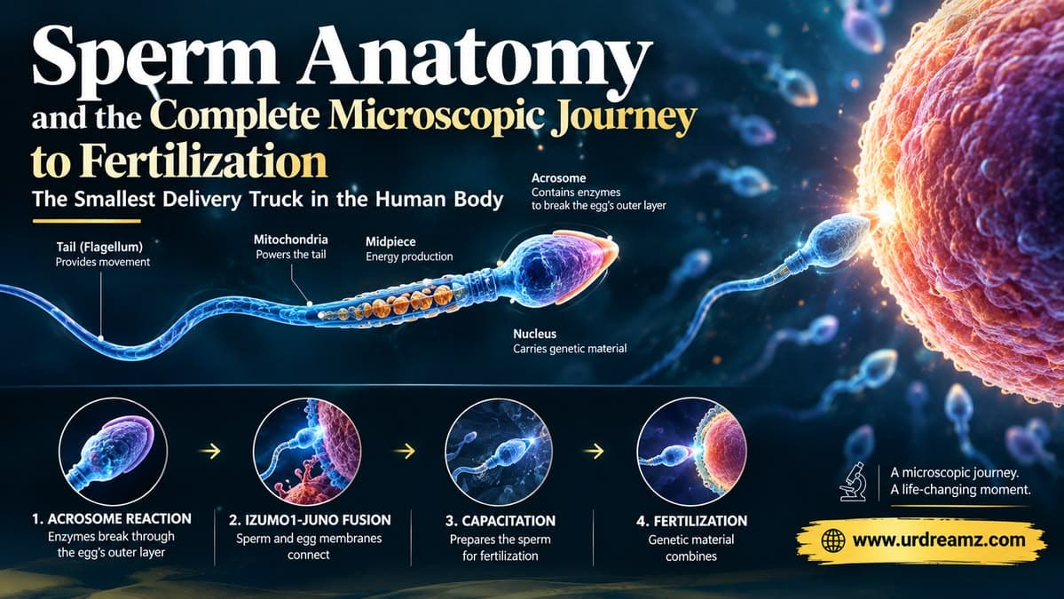
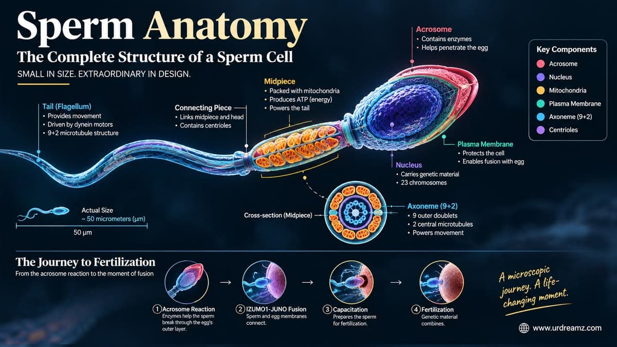
A human sperm cell has two core functions: acting as a delivery vehicle for its genetic cargo, and operating as a self-contained functional machine capable of reaching and fertilizing an egg. Structurally, it is divided into a head (containing the compacted DNA-filled nucleus and the enzyme-loaded acrosome, protected by the perinuclear theca), a neck (containing the proximal and distal centrioles), a midpiece (packed with 25 to 80 mitochondria generating the ATP energy that powers movement, wrapped around outer dense fibers), and a tail or flagellum (built around a whip-like structure called the axoneme, separated from the midpiece by a ring-like structure called the annulus). During fertilization, the acrosome releases two enzymes — hyaluronidase, which breaks down the egg's outer corona radiata layer, and acrosin, which breaks down the inner zona pellucida layer — after which a sperm-surface protein called IZUMO1 binds to the egg's JUNO receptor, triggering membrane fusion and allowing the sperm's 23 chromosomes to enter the egg, completing fertilization.
1. A Journey Too Small to See: Why You Need an Electron Microscope
Before diving into structure, it's worth appreciating just how small we're actually talking about. A single human sperm cell measures roughly 50 to 60 micrometers in total length — that's about one-twentieth of a millimeter, an object so small it sits entirely beyond the resolving power of the naked eye and even most standard light microscopes used in a typical school laboratory. To see the fine internal architecture of a single sperm cell — its membranes, its internal fibers, its enzyme-packed compartments — scientists rely on electron microscopy, a technique that uses focused beams of electrons rather than visible light to achieve magnification thousands of times greater than what an ordinary microscope can offer. Every detail described in this article, from the acrosome's enzymes to the axoneme's internal fiber arrangement, was mapped using exactly this kind of high-powered imaging.
2. Two Jobs, One Purpose: Delivery Truck and Functional Machinery
At its core, a sperm cell exists to do exactly two things, and every single structure we'll cover in this article exists in service of one or both of these two jobs. First, it functions as a delivery vehicle — its entire structural design is built around the singular task of transporting its genetic cargo, the paternal DNA, from the male reproductive system all the way to a waiting egg. Second, it functions as its own functional machinery — unlike a passive package being carried by an external vehicle, the sperm cell is itself the vehicle, equipped with its own onboard power supply, its own propulsion system, and its own set of molecular tools needed to physically penetrate the egg's protective layers once it arrives. Every structure covered in the sections ahead is, in essence, either part of the "cargo protection and delivery" system, or part of the "engine and toolkit" system — and remarkably, in many cases, the very same structure performs both roles simultaneously.
2b. Before the Journey Begins: A Quick Look at Spermatogenesis
Everything described in this article assumes a fully mature sperm cell, but it's worth briefly understanding how that cell came to exist in the first place. Spermatogenesis, the overall process of sperm production, takes place within the seminiferous tubules of the testes and takes approximately 64 to 72 days from start to finish in humans. It begins with diploid germ cells called spermatogonia, which divide and mature through several distinct stages — spermatocytes, then spermatids — undergoing two rounds of meiotic division along the way to reduce their chromosome count from 46 to the 23 found in a mature sperm cell. The final stage, called spermiogenesis, is where a round, relatively unremarkable spermatid transforms dramatically into the streamlined, highly specialized cell this article has been describing — developing its acrosome, compacting its DNA with protamines, shedding excess cytoplasm, and growing its characteristic tail. This entire transformation happens under the close structural and nutritional support of the Sertoli cells discussed earlier, which guide developing sperm cells through every stage of this remarkable, multi-week process.
3. The Complete Map: Four Regions of a Sperm Cell
Before examining individual components in detail, it helps to have the full map laid out clearly. A mature sperm cell is organized into four broad structural regions, moving from front to back.
| Region | Main Components | Primary Role |
|---|---|---|
| Head | Nucleus (compacted DNA), Acrosome, Perinuclear Theca | Carries and protects the genetic cargo; penetrates the egg's outer layers |
| Neck | Proximal and Distal Centrioles | Connects head to tail; contributes to early embryonic cell division |
| Midpiece | Mitochondria, Outer Dense Fibers, Mitochondrial Sheath | Generates the energy (ATP) that powers tail movement |
| Tail (Flagellum) | Axoneme, Annulus, Outer Dense Fibers (extending from midpiece) | Provides propulsion, driving the sperm forward |
4. The Head: Nature's Most Efficient Delivery Capsule
The sperm's head is where the entire mission's cargo is held — the compacted paternal DNA that will combine with the egg's DNA to form a new, complete genetic blueprint. But the head isn't just a passive container; it's equipped, right at its very front, with a specialized structure that functions almost like a protective helmet designed specifically for penetration — the acrosome. This cap-like structure sits over the front two-thirds of the nucleus and houses a concentrated set of enzymes whose entire purpose is to help the sperm physically break through the protective layers surrounding the egg, which we'll examine closely in the sections ahead.
5. Inside the Acrosome: Meet Hyaluronidase and Acrosin
The acrosome carries two principal enzymes, each with a specific, sequential job to do once the sperm finally reaches the egg.
| Enzyme | Target | Function |
|---|---|---|
| Hyaluronidase | Corona Radiata (outer egg layer) | Breaks down the hyaluronic acid-rich matrix holding the outer layer of cells together, clearing a path through it |
| Acrosin | Zona Pellucida (inner egg layer) | A protease enzyme that digests through the glycoprotein-based inner layer surrounding the egg |
These two enzymes don't act simultaneously in a single burst — they function sequentially, corresponding to the two separate protective layers the sperm must pass through, one after another, before ever reaching the egg's own cell membrane.
6. The Egg's Two Protective Layers: Corona Radiata and Zona Pellucida
Just as the sperm arrives equipped with specific tools, the egg arrives protected by two distinct defensive layers, arranged from outside in.
| Layer | Position | Composition |
|---|---|---|
| Corona Radiata | Outermost layer | A layer of follicle cells held together in a hyaluronic acid-rich matrix |
| Zona Pellucida | Inner layer, directly surrounding the egg's cell membrane | A glycoprotein-based structural layer, tougher and more resistant than the outer layer |
Only after both of these layers have been successfully breached does the sperm reach the egg's actual outer membrane, where the final, decisive step of fertilization takes place.
7. Breaking Through: How the Acrosome Reaction Actually Works
The process by which the acrosome releases its enzymatic contents is called the acrosome reaction. As the sperm makes contact with the corona radiata, the outer acrosomal membrane fuses with the sperm's own plasma membrane at multiple points, causing the membrane to essentially open up and release its stored enzymes directly onto the surrounding layers. Hyaluronidase is released first, breaking down the corona radiata's matrix and allowing the sperm to work its way through the outer layer of cells. Once through, acrosin — released as the sperm makes contact with the zona pellucida — takes over, digesting a path through this tougher, deeper barrier. By the time both enzymes have completed their work, the acrosome itself has been largely spent and shed, and what remains of the sperm head is now in direct position to meet the egg's own cell membrane.
Corona Radiata
Released
Cleared
Released
Penetrated
8. IZUMO1: The Key That Finally Opens the Door
Having cleared both of the egg's protective layers, the sperm now faces one final challenge: the egg's own cell membrane does not simply allow any arriving cell to merge with it. This final, critical step depends on a specific protein located on the sperm's head membrane, called IZUMO1. Without IZUMO1 present and functioning correctly, a sperm cell — even one that has successfully broken through both outer layers — cannot actually fuse with the egg at all. This protein acts almost precisely like a key, one that must fit a very specific lock waiting on the egg's own surface in order for the final union to take place.
9. JUNO: The Egg's Side of the Handshake
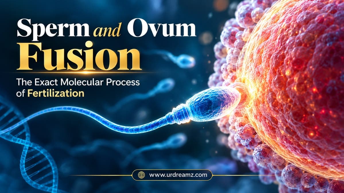
That specific "lock" on the egg's surface is a receptor protein called JUNO. When IZUMO1, on the sperm's surface, binds successfully to JUNO, on the egg's surface, this molecular handshake triggers the actual fusion of the sperm and egg cell membranes — the true, defining moment of fertilization at the cellular level. This IZUMO1-JUNO interaction is now understood to be an essential, non-negotiable step in human fertilization; without a successful bind between these two specific proteins, sperm and egg simply cannot merge into one cell, regardless of how successfully the sperm navigated everything that came before it.
10. The Moment of Fusion: 23 Chromosomes Cross the Threshold
Once IZUMO1 and JUNO bind and the two cell membranes fuse, the sperm's genetic material — 23 chromosomes, exactly half the number found in a typical human cell — enters the egg, which itself carries 23 chromosomes of its own. Together, these two sets combine to form the complete, 46-chromosome genetic blueprint of a brand-new individual. This is the precise cellular moment fertilization is considered complete — the culmination of the entire journey this article has been tracing, from an invisibly small cell equipped with a helmet, two enzymes, and a single key-shaped protein, to the successful, molecular-level creation of a genetically new human blueprint.
10b. Why Only One Sperm Gets In: The Block to Polyspermy
Given that roughly 300 million sperm begin the journey, and typically several hundred actually reach the vicinity of the egg, a natural question arises: what stops more than one sperm from fertilizing the same egg? The answer lies in a rapid, elegant safeguard called the block to polyspermy, which activates within moments of the first successful sperm-egg fusion. The instant one sperm successfully fuses via the IZUMO1-JUNO interaction described earlier, the egg releases the contents of specialized structures called cortical granules, positioned just beneath its outer membrane. These granules release enzymes that rapidly modify the zona pellucida's structure, hardening it and rendering it resistant to any further sperm penetration within seconds. This fast biochemical "lockdown" is critical, since fertilization by more than one sperm — a condition called polyspermy — would result in an embryo with an abnormal, non-viable chromosome count, incompatible with normal development.
11. Traveling Light: Why the Sperm Sheds Its Own Cytoplasm
Here's a genuinely fascinating detail about sperm development that most people never learn: as a sperm cell matures, it deliberately discards much of its own internal cellular machinery — ribosomes, endoplasmic reticulum, and a significant portion of its cytoplasm — material that virtually every other cell in the human body retains and actively uses. This isn't damage or malfunction; it's a deliberate, purposeful stripping-down process. A developing sperm cell essentially "decides" it doesn't need this extra internal machinery for its singular mission, and shedding it makes the final cell lighter, more streamlined, and more efficient for the swimming journey ahead — much like a delivery vehicle being stripped of every unnecessary component to maximize speed and efficiency for one specific, singular task.
12. Sertoli Cells: The Body's Quiet Cleanup Crew
This discarded material doesn't simply float away unused — it's carefully collected and consumed by specialized support cells within the testes called Sertoli cells, sometimes referred to as "nurse cells" because of the supportive role they play throughout the entire sperm development process. As the developing sperm cell sheds its excess cytoplasm — collected into a structure called a residual body, sometimes persisting briefly afterward as a smaller cytoplasmic droplet attached near the midpiece — nearby Sertoli cells absorb and break down this material through a process called phagocytosis, essentially "eating" the leftover cellular material the maturing sperm no longer needs. This tidy, efficient cleanup process ensures nothing goes to waste and that the final, mature sperm cell remains as streamlined and functionally efficient as possible.
13. DNA Packaging Like No Other Cell: Protamines vs. Histones
Nearly every other cell in the human body packages its DNA around proteins called histones — a system that allows DNA to be organized while still remaining accessible for the cell's ongoing, everyday functions like gene expression. Sperm cells are a dramatic exception. During the final stages of sperm development, histones are almost entirely replaced by a different class of proteins called protamines, which are considerably smaller and far more effective at tightly compacting DNA.
| Feature | Histones (Most Cells) | Protamines (Sperm Cells) |
|---|---|---|
| Primary Role | Organize DNA while keeping genes accessible | Compact DNA extremely tightly for transport and protection |
| Compaction Level | Moderate | Extremely high — significantly tighter than histone-based packaging |
| Gene Accessibility | Genes remain accessible for regular cellular activity | DNA is largely inactive/silenced during this compacted state |
| Additional Stabilization | Not typically required | Stabilized further by disulfide bridges |
This switch makes biological sense once you consider what the sperm's DNA actually needs during this specific journey: not active use, but maximum protection during transport through a genuinely hostile physical environment.
14. The Disulfide Bridges That Lock the Genome Tight
Protamines don't just compact DNA on their own — they're further reinforced by chemical bonds called disulfide bridges, which form cross-links between neighboring protamine molecules, essentially locking the entire compacted structure together into an exceptionally stable, tightly bound package. This additional layer of chemical reinforcement is precisely why sperm DNA can survive being tightly packed, physically transported, and exposed to a genuinely challenging journey through the female reproductive tract without the genetic material becoming damaged, tangled, or degraded along the way. Without this specific packaging system, the DNA carried inside each sperm cell would be considerably more vulnerable to exactly the kind of physical and chemical stress this journey inevitably involves.
14b. Not a Complete Swap: Some Histones Remain
It's worth adding an important nuance to the histone-to-protamine story covered earlier: the replacement isn't actually total. In humans, roughly 85 to 95 percent of histones are replaced by protamines during the final stages of sperm maturation, but a small remaining fraction — typically estimated at around 5 to 15 percent — stays in place at specific locations throughout the genome. Research suggests these retained histone regions aren't random leftovers, but may mark genetically important locations, including genes relevant to very early embryonic development, effectively leaving a kind of molecular "bookmark" that becomes relevant again only after fertilization, once the embryo begins actively using its own genetic instructions. This detail adds yet another layer of precision to the picture this article has been building throughout: even the seemingly simple story of "protamines replace histones" turns out, on closer inspection, to be a carefully calibrated, selectively incomplete process, rather than a blunt, all-or-nothing swap.
15. When Packaging Goes Wrong: Sperm DNA Fragmentation and Infertility
This is a genuinely important point for anyone dealing with unexplained fertility difficulties to understand. During spermatogenesis — the overall process by which sperm cells develop — the chromatin (the DNA-protein complex) occasionally fails to remodel and compact correctly. When this happens, the resulting sperm cell can carry what's known as fragmented DNA — meaning the genetic material inside is broken into pieces rather than properly organized and intact. A sperm cell with significantly fragmented DNA may still, in some cases, appear structurally normal and even swim normally, yet be functionally compromised at the genetic level in a way that standard visual examination alone cannot detect. This specific issue is increasingly recognized as a meaningful, underlying contributor in cases of unexplained infertility or recurrent pregnancy loss, and specialized sperm DNA fragmentation testing, distinct from a standard semen analysis, can help identify it when routine fertility investigations don't reveal an obvious cause.
15b. How Doctors Actually Measure Sperm Health: Semen Analysis Basics
Given how much of this article has focused on the sperm cell's internal structure, it's worth briefly covering how doctors actually evaluate sperm health in practice. A standard semen analysis, guided by World Health Organization reference values, typically examines several key parameters together, since no single measurement alone tells the complete story of fertility potential.
| Parameter | What It Measures |
|---|---|
| Sperm Concentration | Number of sperm per milliliter of semen |
| Total Motility | Percentage of sperm that are moving at all |
| Progressive Motility | Percentage of sperm swimming effectively in a forward direction |
| Morphology | Percentage of sperm with a normal, properly formed head, midpiece, and tail shape |
| Volume | Total amount of semen produced per ejaculation |
| DNA Fragmentation Index (specialized test) | Proportion of sperm carrying damaged or improperly packaged DNA, as discussed earlier in this article |
It's worth noting that a sperm cell can appear entirely normal under a standard microscope, with typical motility and shape, while still carrying the kind of fragmented DNA discussed earlier — which is precisely why specialized DNA fragmentation testing exists as a separate, additional evaluation for cases where standard semen analysis results don't fully explain ongoing fertility difficulties.
16. The Perinuclear Theca: A Helmet's Inner Padding
Sitting just beneath the acrosome and surrounding much of the compacted nucleus is a structural protein layer called the perinuclear theca (often abbreviated PT). This cytoskeletal structure functions much like the internal padding inside a protective helmet — it's not the outer shell itself, but a supportive structural layer that helps the head maintain its shape and resist deformation under mechanical stress, particularly during the physically demanding passage through the female reproductive tract. Without this reinforcing layer, the compacted, tightly packaged nucleus we discussed in the previous sections would be considerably more vulnerable to physical damage during the journey ahead.
17. Moving to the Neck: Centrioles, Proximal and Distal
Just behind the head sits the sperm's neck region, home to two small but functionally important structures called centrioles — specifically, a proximal centriole and a distal centriole. The proximal centriole plays a genuinely crucial role after fertilization: it contributes directly to the first cell division of the newly formed embryo, since the egg itself typically lacks a functional centriole of its own at this stage, making the sperm's contribution here essential to normal early embryonic development, not just to delivering DNA. The distal centriole, meanwhile, plays a more structural role, contributing to the organization and support of the flagellum, the whip-like tail structure that powers the sperm's entire forward movement.
18. The Midpiece: Sperm's Own Power Plant
Directly behind the neck sits the midpiece — and if the head is the sperm's cargo compartment, the midpiece is unmistakably its engine room. This section is densely packed with mitochondria, the cell's energy-generating structures, arranged in a tight, helical sheath wrapped around the sperm's central fiber core. This concentrated mitochondrial arrangement exists for one clear reason: to generate, as efficiently as possible, the enormous and continuous energy supply needed to power the tail's relentless beating motion across an genuinely demanding, extended journey.
19. Mitochondria Count and the Energy Behind Every Beat
A single human sperm cell typically contains somewhere between 25 and 80 mitochondria, tightly wound around the midpiece's central fiber structure. These mitochondria continuously generate ATP — adenosine triphosphate, the universal energy currency used by cells throughout the body — and this ATP is transported both forward toward the head and backward along the length of the tail, fueling the sustained, rhythmic beating motion that drives the sperm's swimming movement across its entire journey.
*Illustrative range based on general reproductive biology literature; exact counts vary between individuals.
19b. How Sperm Compares to Every Other Cell in Your Body
It's genuinely worth pausing to appreciate just how different a sperm cell is from virtually every other cell type the human body produces. Most human cells are roughly spherical or irregularly shaped, remain fixed in place within tissue, contain 46 chromosomes, and maintain a full, standard set of internal organelles for ongoing metabolic activity. A sperm cell breaks from this pattern in almost every respect: it's elongated and streamlined rather than rounded, it actively swims rather than staying fixed in place, it carries only 23 chromosomes rather than the usual 46, and, as covered earlier in this article, it deliberately sheds much of its standard cellular machinery rather than retaining it. Few, if any, other cells in the human body are quite this specialized, quite this stripped-down, or quite this singularly focused on one specific, time-limited mission.
20. The Outer Dense Fibers: A Shock Absorber for the Tail
Running alongside the sperm's central fiber core, extending from the neck through the midpiece and into the principal piece of the tail, are nine structures called outer dense fibers, often abbreviated ODF. These fibers act as a kind of shock absorber and structural reinforcement system, helping the tail withstand the mechanical stress of constant, rapid beating without breaking down or losing its structural integrity over the course of the journey. Without this reinforcement, the tail's continuous whip-like motion — repeated many times per second, over an extended distance — would place far more physical strain on the structure than it could realistically withstand intact.
21. The Annulus: A Gatekeeper Between Midpiece and Tail
At the boundary between the midpiece and the principal piece of the tail sits a ring-like structure called the annulus. Its primary function is compartmentalization — essentially acting as a structural gatekeeper that helps keep the mitochondria-rich midpiece region properly separated and organized from the rest of the tail, preventing components from migrating out of place along the length of the flagellum. This might sound like a minor structural detail, but proper compartmentalization at this junction plays a meaningful role in maintaining the sperm's overall structural integrity and function throughout its journey.
22. The Axoneme: The Engine That Powers Movement
Running through the entire length of the tail, from the neck all the way to its very tip, is the axoneme — the actual internal engine responsible for the sperm's characteristic whip-like, beating movement. Structurally, the axoneme is built from a highly organized arrangement of microtubules, and its coordinated, rhythmic bending motion, powered by the ATP generated in the midpiece's mitochondria, is what physically propels the sperm cell forward through fluid. Every visible movement of the tail — every beat, every wave-like undulation driving the sperm onward — ultimately originates from this precisely organized internal structure.
22b. The Tail's Three Sub-Regions: Midpiece, Principal Piece, and End Piece
While this article has already covered the midpiece as its own major region, it's worth noting that the tail itself, running from the annulus to its very tip, is further divided into two additional sub-regions with distinct structural characteristics. The principal piece, making up the majority of the tail's total length, contains the axoneme surrounded by the outer dense fibers discussed earlier, wrapped in a supportive fibrous sheath that provides additional structural rigidity along this longer stretch. The end piece, the very final, tapering segment at the extreme tip of the tail, contains only the bare axoneme itself, without the surrounding outer dense fibers or fibrous sheath present in the principal piece — a deliberately simplified, more flexible design at the very tip, where the tail's motion becomes most whip-like and the sperm's forward-driving force is ultimately generated.
23. Capacitation: The Final Transformation Inside the Female Body
One final, essential transformation takes place only after the sperm has entered the female reproductive tract, called capacitation. During this process, the sperm's outer membrane undergoes a series of biochemical changes — including the removal and rearrangement of certain surface proteins and cholesterol molecules that had been present since ejaculation — essentially priming the sperm for the acrosome reaction described earlier in this article. A sperm cell that hasn't undergone capacitation is not yet capable of successfully binding to and penetrating an egg, even if it physically reaches one; this final biochemical "unlocking" step is a mandatory prerequisite for successful fertilization, ensuring the sperm is only fully "activated" at the appropriate location and time within the female reproductive system, rather than prematurely.
23b. Hyperactivation: Switching Into a More Powerful Swimming Mode
Alongside capacitation, sperm cells undergo a related but distinct change called hyperactivated motility as they approach the egg. In this mode, the tail's beating pattern shifts from the relatively smooth, symmetrical motion used during the earlier part of the journey to a far more forceful, whip-like, asymmetrical beating pattern, generating significantly greater propulsive force. This more powerful swimming mode is thought to be specifically important for helping the sperm physically push through the thick, viscous zona pellucida layer discussed earlier, and for helping it navigate the mucus-rich environment of the female reproductive tract closer to the egg, where a gentler, more energy-efficient swimming style would no longer be sufficient for the final, most physically demanding stretch of the journey.
24. Putting It All Together: The Complete Journey, Step by Step
| Step | What Happens |
|---|---|
| 1. Ejaculation | Roughly 300 million sperm are released |
| 2. Travel Through Female Reproductive Tract | Sperm undergo capacitation, priming them for fertilization |
| 3. Contact With Corona Radiata | Hyaluronidase is released via the acrosome reaction, clearing the outer layer |
| 4. Contact With Zona Pellucida | Acrosin digests through the inner protective layer |
| 5. Membrane Fusion | IZUMO1 on the sperm binds to JUNO on the egg, triggering fusion |
| 6. Genetic Union | 23 sperm chromosomes combine with 23 egg chromosomes, forming a complete 46-chromosome set |
24b. Size Matters: Sperm and Egg in Perspective
It's worth visualizing just how mismatched in size the sperm and egg actually are, since the numbers are genuinely striking. A human egg (oocyte) is roughly 120 micrometers in diameter, making it the largest single cell in the human body and, remarkably, just barely visible to the naked eye under the right conditions — unlike the sperm cell, which requires magnification to see at all. A sperm cell's head alone is only about 4 to 5 micrometers long, meaning the egg is dramatically larger than even the sperm's entire body, let alone just its head.
*Approximate values from standard reproductive biology references; not drawn to a linear scale for visual clarity.
This size mismatch is part of why the egg's two protective layers, the corona radiata and zona pellucida discussed earlier, are so structurally significant relative to the sperm approaching them — from the sperm's perspective, penetrating these layers is genuinely a comparatively massive physical undertaking.
24c. Common Myths About Sperm, Corrected
Given how much misinformation circulates around this topic, a few specific myths deserve direct correction, grounded in everything covered throughout this article. One common myth is that sperm are essentially simple, identical "swimmers" with no meaningful internal complexity — as this entire article has demonstrated, a single sperm cell is an extraordinarily specialized, multi-component structure, with dedicated enzymatic, structural, and energy-generating systems working in precise coordination. Another myth is that a sperm's DNA is simply "along for the ride," passively transported without any active protective mechanism — in reality, as covered in the sections on protamines and disulfide bridges, the genetic material is actively, deliberately compacted and chemically reinforced specifically to survive the journey intact. A third myth treats semen volume or visible thickness as a meaningful indicator of fertility on its own — in clinical reality, fertility potential depends on a combination of factors covered in the semen analysis section, including concentration, motility, morphology, and DNA integrity, none of which can be reliably judged by visual appearance alone.
24d. A Hostile Environment: What Sperm Survive Along the Way
It's worth appreciating just how physically demanding the journey through the female reproductive tract actually is, since it puts the structural reinforcements covered throughout this article into genuine context. The vaginal environment is naturally acidic, a defense mechanism against harmful bacteria, but also a genuinely hostile environment for sperm survival, meaning a significant proportion of the roughly 300 million sperm released never make it far past this initial barrier at all. Those that do survive must then navigate cervical mucus, whose consistency changes throughout the menstrual cycle and is only truly passable around the time of ovulation, followed by a long swim through the uterus and into the fallopian tubes, the actual site where fertilization typically occurs. Of the original hundreds of millions released, only a tiny fraction — often just a few hundred — ever reach the vicinity of the egg at all, which makes every structural safeguard covered in this article, from the perinuclear theca's protective padding to the protamine-packed, disulfide-reinforced DNA, considerably easier to appreciate as genuinely necessary survival features rather than excessive engineering.
24e. How Long Does the Whole Journey Actually Take?
Pulling the timeline together from earlier sections of this article: sperm can typically survive within the female reproductive tract for up to about five days under favorable conditions, though the egg itself remains viable for fertilization for a much shorter window, generally around 12 to 24 hours after ovulation. The physical swim from the point of ejaculation to the fallopian tubes, where fertilization typically takes place, can occur remarkably quickly for the fastest-moving sperm, sometimes within as little as 30 minutes to a few hours, though the full capacitation process discussed earlier continues to prepare sperm for fertilization over a longer window of several hours after entering the reproductive tract. This means successful fertilization depends on a fairly narrow, precisely timed overlap between a surviving, capacitated sperm cell and a viable egg — one more layer of biological precision stacked on top of everything else this article has covered.
25. Why This Design Matters: A Note on Precision
Stepping back from the individual mechanics, what's genuinely remarkable about everything covered in this article is how little of it is arbitrary. Every structure discussed — the enzyme-loaded acrosome, the tightly wound mitochondrial sheath, the protamine-packed nucleus, the shock-absorbing outer dense fibers — exists because it solves a specific, precise problem the sperm cell must overcome on its journey. Nothing here is oversized or undersized for its task; every component is calibrated, down to the molecular level, for exactly the role it needs to perform. For a structure this small — smaller than the width of a human hair, invisible without specialized instruments — to be organized with this degree of functional precision is a genuinely humbling thing to sit with, a reminder of just how deliberately even the smallest, most hidden details of creation appear to have been measured out.
26. Frequently Asked Questions
What are the main parts of a sperm cell?
A sperm cell has four main regions: the head (containing the nucleus and acrosome), the neck (containing centrioles), the midpiece (containing mitochondria), and the tail or flagellum (containing the axoneme).
What is the function of the acrosome?
The acrosome is a cap-like structure at the front of the sperm head that contains enzymes, primarily hyaluronidase and acrosin, which help the sperm penetrate the egg's protective outer layers.
What is the difference between the corona radiata and the zona pellucida?
The corona radiata is the outermost layer of cells surrounding the egg, broken down by hyaluronidase, while the zona pellucida is a tougher, inner glycoprotein layer, broken down by acrosin.
What is IZUMO1 and why is it important?
IZUMO1 is a protein on the sperm's surface that binds to the JUNO receptor on the egg's surface, triggering the fusion of sperm and egg cell membranes, a necessary step for fertilization.
Why does sperm DNA use protamines instead of histones?
Protamines compact DNA far more tightly than histones, providing better protection for the genetic material during the sperm's journey, further stabilized by disulfide bridges.
What causes sperm DNA fragmentation?
It occurs when chromatin fails to remodel and compact correctly during spermatogenesis, resulting in sperm carrying broken or improperly packaged DNA, which can contribute to infertility.
How many mitochondria does a sperm cell have?
A single sperm cell typically contains between 25 and 80 mitochondria, arranged in the midpiece, generating the energy needed to power tail movement.
What is capacitation?
Capacitation is a biochemical transformation sperm undergo inside the female reproductive tract, priming them for the acrosome reaction and making them capable of fertilizing an egg.
What is the axoneme?
The axoneme is the internal microtubule-based structure running through the sperm's tail that powers its whip-like, beating movement.
Why does a maturing sperm cell shed its cytoplasm?
Shedding excess cytoplasm, ribosomes, and other cellular machinery makes the sperm lighter and more streamlined for swimming; the discarded material is absorbed by Sertoli cells through phagocytosis.
What prevents more than one sperm from fertilizing the same egg?
A rapid process called the block to polyspermy hardens the zona pellucida within seconds of the first successful sperm-egg fusion, preventing additional sperm from penetrating.
How many sperm are released in a single ejaculation?
Typically around 200 to 300 million sperm are released, though only a small fraction, often just a few hundred, ever reach the vicinity of the egg.
How big is a human egg compared to a sperm cell?
A human egg is roughly 120 micrometers in diameter, making it the largest cell in the human body, while a sperm head is only about 4 to 5 micrometers long.
What is spermatogenesis?
Spermatogenesis is the overall process of sperm production in the testes, taking about 64 to 72 days from initial germ cells to a fully mature sperm cell.
What is hyperactivated motility?
It's a more forceful, whip-like swimming pattern sperm adopt as they approach the egg, generating greater propulsive force to help penetrate the zona pellucida.