Chronic Stomach Pain? Know About H. pylori — The Bacteria Living in Half the World's Stomachs
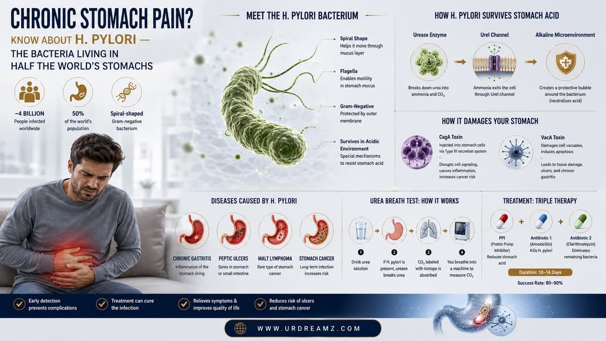
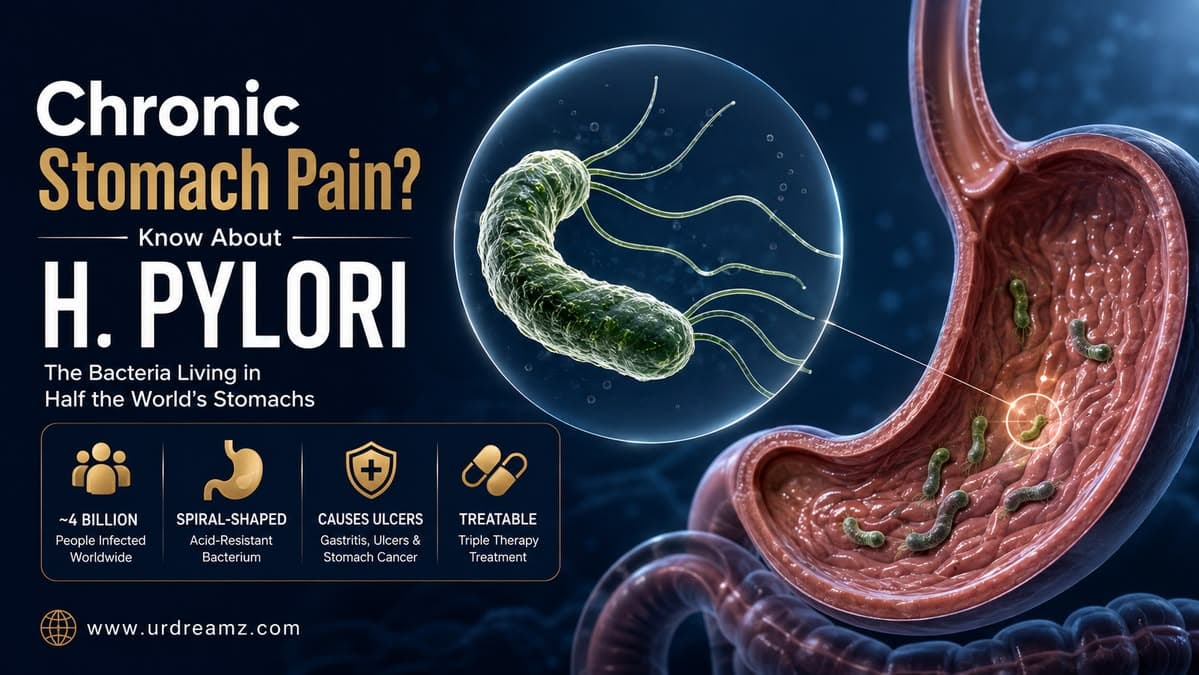
Somewhere between 4 and 4.4 billion people on Earth — roughly half the world's population — are currently carrying a spiral-shaped bacterium in their stomach called Helicobacter pylori (H. pylori). Most carriers never know it's there. Yet this single organism has solved one of the strangest survival puzzles in all of microbiology: how to live permanently inside an organ filled with acid strong enough to dissolve metal. This guide explains exactly how H. pylori pulls this off, why it causes serious harm in only some people while leaving others completely unaffected, how doctors actually test for it using a breath sample, and what modern treatment looks like.
Along the way, this guide walks through the entire biological story in order: how the bacterium physically reaches and anchors itself to the stomach lining, the elegant chemical trick it uses to survive an acid bath that would destroy nearly any other organism, the two toxins responsible for most of the serious damage it can cause, the progression from mild inflammation to ulcers and, in rare cases, cancer, and finally, exactly how modern medicine detects and treats this infection.
1. What Is H. pylori?
Helicobacter pylori, commonly abbreviated as H. pylori, is a spiral-shaped, gram-negative bacterium that colonizes the human stomach. Its distinctive corkscrew shape is not just a curiosity — it is directly functional, allowing the bacterium to physically burrow and move through the thick, viscous mucus layer that coats the inside of the stomach. Being classified as gram-negative refers to a specific structural feature of its outer cell wall, a classification detail that matters clinically because it influences which antibiotics are effective against it.
2. How Common Is H. pylori Infection Worldwide?
Out of a global population of roughly 8 billion people, somewhere between 45% and 50% are estimated to carry H. pylori in their stomach — translating to approximately 4 billion people worldwide currently living with this bacterium. This makes H. pylori one of the most widespread bacterial infections on the planet, though prevalence varies significantly by region, generally being far higher in developing countries and regions with limited access to clean water and sanitation infrastructure, and comparatively lower in many developed countries with modern hygiene standards.
3. Why Doctors Recommend H. pylori Testing for Stomach Pain
When a patient presents with persistent or recurring stomach pain, doctors frequently recommend H. pylori testing as part of the diagnostic workup, since this bacterium is one of the most common identifiable causes of chronic gastric discomfort, gastritis, and peptic ulcers. Identifying whether H. pylori is present helps doctors distinguish between stomach pain caused by this treatable bacterial infection versus pain arising from other causes, such as dietary factors, stress-related acid imbalance, or other gastrointestinal conditions entirely unrelated to bacterial colonization.
4. Understanding the Stomach's Protective Mucosal Lining
The inner surface of the stomach is lined with a single layer of specialized cells called epithelial cells, which form a protective barrier between the stomach's harsh internal contents and the deeper tissue layers beneath. Covering this epithelial layer is a thick, gel-like layer of mucus known as the mucosal lining. This mucosal layer serves a critical protective function for the body, acting as a physical barrier and buffer that helps shield the underlying epithelial cells from being directly damaged by the stomach's own powerful digestive acid.
5. How H. pylori Reaches the Epithelial Cells
When H. pylori bacteria enter the stomach, they do not remain suspended in the highly acidic central cavity of the stomach, where survival would be extremely difficult even briefly. Instead, using their characteristic spiral shape and whip-like flagella for propulsion, they actively burrow down through the thick mucosal layer, working their way toward the epithelial cell surface underneath. Once they reach this deeper location, they effectively hide themselves beneath the protective mucus layer, positioning themselves in a location that is both closer to a stable nutrient supply and comparatively shielded from the harshest acid concentrations found in the open stomach cavity.
6. BabA and SabA: The Anchoring Proteins
To successfully establish itself at the epithelial cell surface, H. pylori relies on specialized surface proteins called BabA and SabA, often referred to as anchoring proteins. These proteins function like molecular grappling hooks, allowing the bacterium to physically bind and attach itself firmly to specific receptor sites on the surface of epithelial cells. This anchoring mechanism is essential for long-term colonization, since without a firm attachment, the bacterium would risk being swept away along with the stomach's natural mucus turnover and emptying processes.
BabA specifically recognizes and binds to a particular sugar-based marker displayed on the surface of certain epithelial cells, a highly specific lock-and-key interaction that helps explain why H. pylori shows such a strong preference for colonizing the stomach lining rather than other tissues throughout the digestive tract. SabA works in a broadly similar way, recognizing a different but related surface marker that becomes more prominent on epithelial cells during periods of ongoing inflammation, meaning SabA-mediated attachment can actually become stronger the longer an infection persists and the more inflamed the local tissue becomes — a self-reinforcing cycle that helps explain why chronic H. pylori infections can be so difficult for the body to clear entirely on its own, without medical treatment.
7. The Real Puzzle: How Does H. pylori Survive Stomach Acid?
The central biological mystery surrounding H. pylori has always been straightforward to state but remarkable to explain: the human stomach contains hydrochloric acid (HCl), a substance strong enough to break down food and kill the vast majority of bacteria that enter the digestive tract. So how does H. pylori not only survive, but thrive, inside this hostile acidic environment for years or even decades? The answer lies in one of the most elegant survival mechanisms found anywhere in microbiology — a self-generated, localized neutralization system built around a single enzyme and a naturally occurring waste compound already present in the human body.
To put the scale of this challenge in perspective, the stomach's resting pH typically sits somewhere between 1.5 and 3.5 — an acidity level comparable to battery acid or lemon juice, and roughly a million times more acidic than neutral water. Almost no bacteria are capable of surviving direct, prolonged exposure to an environment this harsh, which is precisely why the stomach has historically been considered one of the most naturally sterile organs in the human body. H. pylori's ability to not just survive but establish a permanent, thriving colony under these conditions is what first drew intense scientific curiosity to this organism after its discovery in the 1980s, eventually earning its discoverers a Nobel Prize for overturning decades of medical assumptions about the causes of stomach ulcers.
8. Where Urea Comes From: A Journey From the Liver
To understand H. pylori's survival trick, it helps to first understand where urea comes from in the human body. The liver continuously processes waste products generated from normal metabolism, and one of its key jobs is converting toxic ammonia byproducts into a much safer compound called urea, which is the very same waste substance eventually excreted from the body in urine. This urea does not stay confined to the kidneys and urinary system — it circulates throughout the bloodstream and, in the process, some of it naturally passes through and reaches the stomach lining as part of normal blood circulation to the digestive tract.
9. UreI: The pH-Gated Channel
H. pylori possesses a specialized channel embedded in its outer membrane called UreI. Under normal, non-acidic conditions, this channel remains closed, effectively acting as a locked door. However, when the surrounding environment becomes acidic — precisely the condition H. pylori encounters in the stomach — this UreI channel opens in response, allowing circulating urea from the surrounding tissue to flow directly into the bacterium's interior. This pH-sensitive gating mechanism is a remarkably efficient design: the channel only opens exactly when the bacterium needs it most, conserving cellular resources during less acidic conditions.
10. Urease: The Enzyme That Changes Everything
Once urea has entered the bacterium through the open UreI channel, it encounters an enzyme called urease, which H. pylori produces in remarkably large quantities compared to most other bacteria — urease is, in fact, one of the most abundant proteins found in H. pylori's cellular makeup. This enzyme is responsible for breaking down, or metabolizing, the urea molecule into simpler byproducts, setting off the chemical chain reaction that ultimately allows H. pylori to neutralize the acid immediately surrounding it.
11. From Urea to Ammonia: Breaking Down the Chemistry
When urease breaks down urea, the reaction produces two byproducts: ammonia (NH3) and carbon dioxide (CO2). The ammonia produced through this reaction is then released outward from the bacterium into the immediately surrounding stomach environment. Because stomach acid is, at its core, a solution rich in hydrogen ions (which is what makes it acidic), the released ammonia reacts directly with these hydrogen ions, combining with them to form ammonium ions (NH4+). This is the critical chemical step: ammonium formation actively consumes and removes free hydrogen ions from the immediate local environment, directly counteracting and neutralizing the acidity in that specific area.
| Step | Process | Result |
|---|---|---|
| 1 | Urea enters bacterium through open UreI channel | Urea available inside bacterium |
| 2 | Urease enzyme breaks down urea | Ammonia (NH3) and Carbon Dioxide (CO2) produced |
| 3 | Ammonia released into surrounding acidic environment | Ammonia reacts with free hydrogen ions |
| 4 | Ammonia combines with hydrogen ions | Ammonium (NH4+) formed; local acidity reduced |
12. Creating a Personal Safe Zone Inside a Sea of Acid
The end result of this urea-to-ammonia-to-ammonium reaction chain is that H. pylori effectively creates a small, localized "safe zone" of neutralized pH directly surrounding its own cell body, even while the broader stomach environment remains intensely acidic just a short distance away. This is not a strategy for neutralizing the entire stomach's acidity — that would be neither necessary nor achievable — but rather a highly targeted, localized defense mechanism that only needs to protect the immediate microenvironment around each individual bacterium. This clever, self-generated buffer zone is precisely what allows H. pylori to survive in a location that would otherwise be lethal to nearly any other bacterial species.
13. Why H. pylori Can Survive for 20+ Years
Because this acid-neutralization mechanism is continuously active and self-sustaining as long as urea remains available from the surrounding blood supply, H. pylori is capable of maintaining a stable, protected colony within the stomach lining for extremely long periods — commonly persisting for 20 years or more, and in many cases, for a person's entire lifetime if left untreated. This chronic persistence is part of what makes H. pylori such a medically significant organism, since even low-level, long-term inflammation caused by its continued presence can eventually contribute to serious health complications over time.
14. Why Some Carriers Never Get Sick
Despite roughly 4 billion people worldwide carrying H. pylori, only a relatively small percentage — commonly cited as somewhere between 10% and 20% of carriers — go on to develop clinically significant disease such as gastritis, peptic ulcers, or gastric cancer. This variability depends on several interacting factors, including the specific strain of H. pylori involved (since not all strains produce the same toxins in equal amounts), individual genetic susceptibility, the strength and behavior of a person's own immune response, and environmental factors such as diet and smoking. This is precisely why some individuals can carry the bacterium for decades with no noticeable symptoms, while others develop significant gastrointestinal disease relatively early after infection.
This distinction matters enormously in clinical practice, since it means a positive H. pylori test result alone does not automatically indicate an urgent medical crisis, nor should it be dismissed as irrelevant. Doctors typically weigh a positive test result together with the patient's actual symptoms, any visible findings from imaging or endoscopy, and relevant risk factors like family history of stomach cancer, before deciding on the most appropriate course of action. In many asymptomatic cases, particularly in regions where H. pylori prevalence is extremely high, physicians may still recommend treatment as a precaution, given the bacterium's well-documented long-term cancer risk, even in the absence of immediate symptoms.
15. The Two Major Toxins: VacA and CagA
The strains of H. pylori most closely associated with serious disease typically produce one or both of two major toxins: VacA (Vacuolating cytotoxin A) and CagA (Cytotoxin-associated gene A). These two toxins work through distinctly different mechanisms to damage the stomach's epithelial cells, and their presence — or absence — in a given H. pylori strain is one of the most significant factors determining whether an infected person is likely to develop serious complications. Not every H. pylori strain circulating in the human population carries the genes needed to produce these toxins, and strains that do carry the CagA gene in particular are consistently associated with substantially higher rates of peptic ulcer disease and gastric cancer in epidemiological studies compared to CagA-negative strains, making toxin profile an important consideration in understanding an individual patient's overall risk.
| Toxin | Primary Mechanism | Effect on Cell |
|---|---|---|
| CagA | Injected directly into the cell's interior | Disrupts internal cell structure and signaling |
| VacA | Acts on the cell's outer membrane | Creates vacuoles (holes), damaging the cell from outside in |
16. CagA: Sabotaging the Cell From Within
The CagA toxin is injected directly into the interior of epithelial cells through a specialized bacterial delivery system. Once inside the cell, CagA interferes with and disrupts the cell's normal internal structural framework and signaling pathways, effectively sabotaging the cell's regular function from the inside. This internal disruption can alter how the cell grows, divides, and communicates with neighboring cells — changes that, over long periods of chronic infection, are believed to contribute meaningfully to the abnormal cellular changes associated with increased cancer risk.
17. VacA: Punching Holes in Cell Membranes
The VacA toxin, by contrast, acts primarily on the outer surface of epithelial cells, creating physical holes, or vacuoles, in the cell membrane. This structural damage to the cell's outer boundary compromises the cell's integrity and normal function, effectively damaging it "from the outside in," in contrast to CagA's more internal, structural sabotage. Together, VacA and CagA represent two complementary strategies through which certain H. pylori strains actively damage the epithelial cells they colonize.
18. How Damaged Cells Trigger Inflammation
Whenever cells in the human body sustain damage — whether from VacA, CagA, or any other harmful influence — they respond by releasing signaling molecules called cytokines. These cytokines serve as distress signals, recruiting immune cells to the site of damage and triggering an inflammatory response intended to address the threat. In the case of chronic H. pylori infection, this cytokine release becomes an ongoing, repeated process, since the bacteria continue producing their toxins and damaging cells over an extended period, resulting in a state of persistent, low-grade inflammation in the stomach lining — a condition that itself becomes clinically significant over time.
Somewhat counterintuitively, this immune response, while intended to protect the body, can itself become part of the problem in chronic H. pylori infection. Because the bacterium is skilled at evading complete clearance by the immune system — partly through the acid-neutralizing safe zone described earlier, and partly through additional immune-evasion strategies — the resulting inflammatory response often becomes a prolonged, unresolved state rather than a short, successful defense. Over years, this chronic inflammatory environment, driven by repeated cytokine signaling, is now understood to be a major contributor to the tissue changes that eventually lead to gastritis, ulcers, and in rare cases, cancer, rather than the direct toxin damage acting entirely alone.
19. From Gastritis to Peptic Ulcer to Gastric Cancer
The disease process associated with harmful strains of H. pylori tends to follow a general, progressive pathway, though not every infected person progresses through every stage. It commonly begins with gastritis, can advance to peptic ulcer disease, and in a smaller subset of long-term, untreated cases, may eventually progress to gastric carcinoma (stomach cancer) — recognized by major health organizations as the most severe potential outcome of chronic H. pylori infection.
It is worth emphasizing that this progression is neither inevitable nor typically rapid — the full pathway from initial infection to gastric cancer, when it does occur, generally unfolds over many years or even decades of sustained, untreated inflammation, rather than happening as a quick or predictable sequence. This extended timeline is actually good news from a public health perspective, since it means there is typically a substantial window of opportunity for detection and treatment at the earlier, more easily managed gastritis or ulcer stages, well before any progression toward more serious disease would even become possible.
| Stage | Description |
|---|---|
| Gastritis | Inflammation of the stomach lining, often an early-stage response to infection |
| Peptic Ulcer | Open sores or wounds developing in the stomach or upper small intestine lining |
| Gastric Carcinoma | Cancer of the stomach; the most serious long-term potential outcome |
20. Understanding Gastritis
Gastritis refers to inflammation of the stomach's inner lining and is typically the earliest visible sign of tissue-level harm caused by H. pylori's toxins and the resulting immune response. Gastritis can present with symptoms such as upper abdominal discomfort, nausea, bloating, or a feeling of fullness after eating only a small amount of food, though some individuals with mild gastritis may experience minimal or no noticeable symptoms at all.
21. Understanding Peptic Ulcers
If chronic inflammation and cellular damage from H. pylori continue unaddressed, the protective lining of the stomach or the upper portion of the small intestine (the duodenum) can eventually break down enough to form an open sore, known as a peptic ulcer. Peptic ulcers are frequently associated with burning or gnawing pain in the upper abdomen, and this pain often has a distinctive relationship with meals — sometimes worsening on an empty stomach and improving temporarily after eating, or in other cases worsening shortly after eating, depending on the ulcer's specific location.
22. How H. pylori Can Trigger Gastric Cancer
In a relatively small subset of chronically infected individuals, particularly those carrying more virulent, CagA-positive strains, decades of persistent inflammation and cellular damage can eventually lead to abnormal changes in the stomach lining's cellular structure. Over a long enough timeline, these abnormal cellular changes can progress toward gastric carcinoma. Because of this well-established link, H. pylori has been formally classified by international health authorities as a recognized risk factor for stomach cancer, making early detection and treatment of the infection a meaningful component of long-term cancer risk reduction for chronically infected individuals.
23. When Stomach Acid Turns Against Its Own Lining
Under normal, healthy conditions, the stomach carefully balances the production of digestive substances like hydrochloric acid (HCl) and the enzyme pepsin against its own protective mucosal defenses, allowing digestion to proceed without self-damage. However, when H. pylori disrupts this balance — through direct toxin damage, chronic inflammation, and interference with normal protective mechanisms — the stomach's own HCl and pepsin can end up acting excessively against the already-weakened epithelial lining. In effect, the digestive substances the body normally relies on for breaking down food end up contributing to further self-inflicted tissue damage once the protective balance has been sufficiently disrupted by the underlying infection.
24. Why Symptoms Are Often Linked to Eating
Many people with H. pylori-related stomach conditions notice that their pain and discomfort has a clear relationship with meals. This connection exists because the stomach releases larger quantities of HCl and pepsin specifically around meal times to support digestion — and if the stomach lining has already been weakened or damaged by chronic H. pylori infection, this natural, meal-triggered surge in acid and digestive enzyme activity can directly aggravate already-compromised tissue, producing noticeable pain that correlates closely with eating patterns.
25. GI Bleeding: A Serious Warning Sign
In more advanced or severe cases of peptic ulcer disease related to H. pylori infection, the damaged tissue can progress to the point of actively bleeding into the gastrointestinal tract. This can present as visible blood in vomit, dark or tar-like stools (a sign of digested blood passing through the digestive tract), or, in less obvious cases, may only be detectable through laboratory testing revealing unexplained anemia. Gastrointestinal bleeding of this kind represents a serious complication requiring prompt medical evaluation, since it can, in severe cases, become a medical emergency.
26. Recognizing the Symptoms of H. pylori Infection
Symptoms of H. pylori infection can range from completely absent to quite severe, depending on the individual and the extent of underlying tissue damage. Common symptoms include burning or gnawing upper abdominal pain, bloating, frequent burping, nausea, loss of appetite, unintentional weight loss, and, in more advanced cases, symptoms of gastrointestinal bleeding as described above. Because many of these symptoms overlap with other, less serious digestive conditions, testing remains the only reliable way to confirm whether H. pylori is actually the underlying cause.
| Symptom | Typical Cause / Stage |
|---|---|
| Burning or gnawing upper abdominal pain | Gastritis or peptic ulcer |
| Bloating and frequent burping | Early gastritis |
| Nausea and loss of appetite | Gastritis or ulcer irritation |
| Unintentional weight loss | More advanced or chronic disease |
| Dark, tar-like stools or vomiting blood | Gastrointestinal bleeding; requires urgent evaluation |
It bears repeating that a genuinely significant proportion of infected individuals experience no symptoms whatsoever, sometimes for their entire lives, which is precisely why H. pylori is often described in medical literature as a largely "silent" infection for most of the people who carry it. This silent nature is also why testing based purely on symptoms will inevitably miss a substantial number of infected individuals, and why some clinicians recommend broader screening in populations known to be at higher risk, such as those with a family history of gastric cancer or those living in high-prevalence regions.
27. Diagnosing H. pylori: An Overview of Testing Methods
Several different diagnostic methods are available to detect H. pylori infection, each with its own advantages, limitations, and appropriate clinical use cases. The most widely used and highly regarded of these is the Urea Breath Test (UBT), though stool antigen testing, blood antibody testing, and endoscopic biopsy are also used depending on the clinical situation.
28. The Urea Breath Test (UBT) Explained Step by Step
The Urea Breath Test works by directly exploiting H. pylori's own urease-based survival mechanism described earlier in this article, turning the bacterium's defense system into a detectable diagnostic signal. This is genuinely one of the more elegant diagnostic tests used in modern medicine, since it does not rely on detecting the bacterium itself directly, but instead detects clear, unambiguous evidence of its unique metabolic activity — an approach that is both highly sensitive and comparatively simple to administer compared to more invasive testing options. The test begins with the patient breathing into an empty collection bag, providing a baseline breath sample before any medicine is given. The patient is then given a special dose of urea in which the normal carbon atom has been swapped out for a specific carbon isotope — either carbon-13 or carbon-14 — rather than the ordinary carbon-12 found in everyday urea.
After swallowing this isotope-labeled urea, the patient waits approximately 20 minutes. If H. pylori is present in the stomach, its abundant urease enzyme will break down this labeled urea exactly as it would with ordinary urea from the bloodstream, producing carbon dioxide (CO2) as one of the reaction byproducts — except in this case, the carbon dioxide produced carries the distinctive carbon-13 or carbon-14 isotope rather than standard carbon-12. The patient then breathes into a second collection bag, capturing this isotope-labeled carbon dioxide if it has been produced.
29. Why Isotopes Make the Urea Breath Test Work
Both breath samples are sent to a laboratory, where specialized equipment — commonly a mass spectrometer or an infrared isotope analyzer — measures and compares the ratio of labeled to unlabeled carbon dioxide present in each sample. If the second breath sample shows a meaningfully elevated presence of carbon-13 or carbon-14 compared to the baseline sample, this confirms that the swallowed urea was broken down inside the stomach by urease — and since human cells do not produce meaningful amounts of urease on their own, a positive result strongly indicates the presence of an active H. pylori infection. This test also includes an antibody-based detection option in some settings, though antibody testing is generally considered less accurate than the isotope-based breath test, since antibodies can remain detectable in the blood long after an infection has already been successfully treated, making the two methods suited to somewhat different clinical purposes.
One important practical consideration with the Urea Breath Test is that certain medications can interfere with its accuracy and must typically be paused for a period beforehand. Proton pump inhibitors, such as omeprazole, and antibiotics can temporarily suppress H. pylori activity enough to produce a falsely negative result, even if the infection is still genuinely present. Because of this, doctors generally instruct patients to stop PPI medications for roughly one to two weeks, and antibiotics or bismuth-containing medications for about four weeks, before undergoing the test, ensuring that any result obtained genuinely reflects the true underlying infection status rather than being distorted by recent medication use.
30. Other Diagnostic Methods: Stool, Blood, and Biopsy
Beyond the Urea Breath Test, several other diagnostic approaches are commonly used in clinical practice, each suited to different situations.
| Test | How It Works | Notes |
|---|---|---|
| Urea Breath Test (UBT) | Detects isotope-labeled CO2 from urease activity in exhaled breath | Highly accurate; commonly used for both diagnosis and confirming successful treatment |
| Stool Antigen Test | Detects H. pylori proteins directly in a stool sample | Non-invasive; also useful for confirming treatment success |
| Blood Antibody Test | Detects antibodies the immune system produced against H. pylori | Less accurate; cannot distinguish current infection from a past, already-treated one |
| Endoscopic Biopsy | Direct tissue sample taken during endoscopy, examined in a lab | Invasive but highly accurate; allows direct visualization of ulcers or tissue damage |
31. Treatment: Understanding Triple Therapy
The standard first-line treatment for confirmed H. pylori infection is known as triple therapy, a treatment regimen combining one acid-suppressing medication with two different antibiotics, taken together for a set course, typically lasting one to two weeks. This combination approach is specifically designed to both create a more hostile environment for the bacteria by reducing stomach acidity and directly kill the bacteria through antibiotic action, working together to clear the infection more effectively than any single medication alone could achieve.
Combining multiple antibiotics simultaneously, rather than relying on a single antibiotic alone, is also a deliberate strategy to reduce the risk of the bacterium developing resistance during treatment. If only one antibiotic were used, any bacteria that happened to carry a chance genetic mutation conferring resistance to that specific drug could survive and multiply, potentially leading to a treatment-resistant infection. By attacking the bacterium simultaneously through two different antibiotic mechanisms, the odds that any individual bacterial cell would happen to carry resistance to both drugs at once are dramatically lower, which is why multi-drug regimens have become the standard approach for treating H. pylori worldwide.
32. The Role of Omeprazole in Treatment
Omeprazole, a medication belonging to a class of drugs called proton pump inhibitors (PPIs), is commonly used as the acid-suppressing component of triple therapy. Omeprazole works by significantly reducing the stomach's production of hydrochloric acid, raising the stomach's overall pH. This serves two important purposes in treatment: it helps the already-damaged stomach lining begin healing by reducing ongoing acid-related irritation, and it also helps improve the effectiveness of the accompanying antibiotics, since certain antibiotics function more reliably in a less acidic environment.
There is also an interesting secondary effect worth understanding: by raising the stomach's pH, omeprazole indirectly places additional pressure on H. pylori itself. Recall that the bacterium's UreI channel and urease system exist specifically to counteract a highly acidic environment; in a stomach where acid has already been substantially reduced by medication, the bacterium's normal defensive advantage becomes considerably less necessary for its own survival, but this same reduced-acid environment simultaneously makes the accompanying antibiotics dramatically more effective, since many antibiotics are known to break down or lose potency when exposed to strongly acidic conditions.
33. The Three Antibiotics Used in Standard Treatment
Alongside omeprazole, standard triple therapy regimens typically include two antibiotics selected from a small group of options known to be effective against H. pylori, commonly including clarithromycin (erythromycin family), amoxicillin, and metronidazole. The specific combination chosen can vary based on regional antibiotic resistance patterns, patient allergies, and prior treatment history.
| Medication | Category | Role in Treatment |
|---|---|---|
| Omeprazole | Proton pump inhibitor (PPI) | Reduces stomach acid production, supports healing |
| Clarithromycin (erythromycin family) | Antibiotic | Directly targets and kills H. pylori bacteria |
| Amoxicillin | Antibiotic | Directly targets and kills H. pylori bacteria |
| Metronidazole | Antibiotic | Directly targets and kills H. pylori bacteria; alternative option in resistant cases |
34. Why Current Antibiotics Have a Fundamental Limitation
From a purely scientific standpoint, current antibiotics used in triple therapy do not directly target H. pylori's remarkable UreI channel and urease-based acid-neutralization system — the very mechanism that allows the bacterium to survive in the stomach in the first place. Instead, they work through more general antibacterial mechanisms. This is a meaningful limitation, because if a treatment could directly block or disable the UreI channel itself, the bacterium would lose its ability to neutralize the surrounding acid and would likely be destroyed by the stomach's own natural acidity almost immediately, without necessarily requiring conventional antibiotics at all.
35. The Future: Targeting the UreI Channel Directly
Recognizing this potential, ongoing scientific research is actively exploring the development of new drugs specifically designed to inhibit the UreI channel directly, preventing it from opening and allowing urea to enter the bacterium in the first place. If such a drug could be successfully developed, it would represent a fundamentally different and potentially more effective therapeutic approach — rather than relying on broad-spectrum antibiotics that can also affect beneficial bacteria elsewhere in the digestive system and contribute to antibiotic resistance over time, a UreI-targeted treatment could specifically and efficiently neutralize H. pylori's core survival mechanism, offering a more precise, mechanism-based path to treatment.
This kind of mechanism-based drug design, sometimes described as targeting a pathogen's specific "virulence factor" rather than simply trying to kill it outright with a broad antibiotic, represents a growing area of interest across infectious disease research generally, not just for H. pylori. The theoretical appeal is significant: because a UreI-blocking drug would exploit a feature entirely unique to H. pylori's own specialized survival strategy, it would be far less likely to disrupt the vast community of other, beneficial bacteria that naturally live throughout the human digestive tract, potentially avoiding many of the broader side effects and resistance concerns associated with current multi-antibiotic treatment regimens. As of now, this approach remains an active area of research rather than an available clinical treatment, meaning triple therapy remains the standard, proven approach for the foreseeable future.
36. Risk Factors: Genetics, Immunity, and Geography
Several factors influence both the likelihood of acquiring H. pylori infection and the likelihood of developing significant disease once infected. Genetic predisposition plays a meaningful role, with some individuals appearing to be genetically more susceptible to H. pylori-related disease than others. A weakened or naturally less robust immune system can also allow the bacterium to cause more significant tissue damage over time compared to individuals with a stronger immune response. Nutrition and overall health status further influence outcomes, since malnutrition and poor overall health can impair the body's ability to repair the ongoing low-grade tissue damage associated with chronic infection. Geography also plays a substantial role — H. pylori prevalence in parts of Asia has been reported at rates exceeding 70% of the population, reflecting a combination of population density, sanitation infrastructure, and historical transmission patterns in the region.
Age at the time of initial infection also appears to matter considerably. In many high-prevalence regions, H. pylori is typically acquired during early childhood, often through close family contact, and this early acquisition means the bacterium has decades to interact with the stomach lining before symptoms, if any, eventually emerge in adulthood. This long latency period is part of why H. pylori-related disease is more commonly diagnosed in middle-aged and older adults, even though the original infection frequently dates back to early childhood, sometimes making it difficult for patients to identify any specific point of transmission.
| Risk Factor | How It Influences H. pylori Outcomes |
|---|---|
| Genetics | Some individuals are genetically more susceptible to related disease |
| Immune system strength | Weaker immune response can allow more significant tissue damage |
| Nutrition and overall health | Poor nutrition can impair tissue repair and healing capacity |
| Geography / sanitation | Prevalence exceeds 70% in parts of Asia; linked to sanitation infrastructure |
| Bacterial strain (CagA/VacA status) | More virulent strains carry significantly higher disease risk |
37. Prevention and Reducing Transmission
H. pylori is generally believed to spread primarily through oral-to-oral or fecal-to-oral transmission routes, meaning it can spread through close personal contact, contaminated food, or contaminated water sources, particularly in areas with inadequate sanitation infrastructure. Practical prevention measures include maintaining good personal hygiene, particularly thorough handwashing before eating and after using the restroom, ensuring access to clean, safe drinking water, and practicing proper food safety and preparation hygiene. While these measures cannot guarantee complete protection given how widespread the bacterium already is globally, they meaningfully reduce the risk of transmission, particularly within households where one family member has already been diagnosed with active infection.
Because H. pylori often spreads within families, particularly from parents to young children, some clinicians recommend that close family members of a newly diagnosed patient consider testing as well, especially if they are also experiencing digestive symptoms. This household-level awareness can be particularly valuable in preventing repeated re-infection after successful treatment, since living in close contact with an untreated, actively infected family member creates an ongoing opportunity for transmission even after a treated individual has successfully cleared their own infection.
38. Frequently Asked Questions
- Q1: What is H. pylori?
- H. pylori (Helicobacter pylori) is a spiral-shaped, gram-negative bacterium that colonizes the human stomach and is one of the most common bacterial infections worldwide.
- Q2: How does H. pylori survive stomach acid?
- H. pylori uses a pH-gated channel called UreI to absorb urea from the bloodstream, then breaks it down using the urease enzyme into ammonia, which neutralizes acid in the bacterium's immediate surroundings.
- Q3: Is H. pylori infection always harmful?
- No. While roughly half the world's population carries H. pylori, only about 10-20% of carriers go on to develop significant disease such as gastritis, peptic ulcers, or gastric cancer.
- Q4: What are VacA and CagA?
- VacA and CagA are two toxins produced by more virulent strains of H. pylori; VacA damages the outer cell membrane, while CagA is injected into cells and disrupts their internal structure and signaling.
- Q5: Can H. pylori cause stomach cancer?
- Yes. Chronic infection, particularly with CagA-positive strains, is a recognized risk factor for gastric carcinoma, though this occurs in only a small subset of long-term, untreated infections.
- Q6: What is the Urea Breath Test?
- The Urea Breath Test (UBT) is a diagnostic test that has a patient swallow isotope-labeled urea; if H. pylori is present, its urease enzyme breaks down the urea and produces detectable labeled carbon dioxide in the breath.
- Q7: What is triple therapy for H. pylori?
- Triple therapy is the standard treatment combining a proton pump inhibitor (such as omeprazole) with two antibiotics, typically taken for one to two weeks to clear the infection.
- Q8: Which antibiotics are used to treat H. pylori?
- Common antibiotics used include clarithromycin, amoxicillin, and metronidazole, usually two of the three combined with omeprazole.
- Q9: Why does H. pylori pain often relate to eating?
- Because the stomach releases more acid and digestive enzymes around meal times, this can aggravate an already-weakened stomach lining damaged by chronic H. pylori infection.
- Q10: How is H. pylori transmitted?
- It is generally believed to spread through oral-to-oral or fecal-to-oral routes, often via contaminated food, water, or close personal contact, particularly in areas with limited sanitation.
- Q11: How common is H. pylori infection?
- Approximately 45-50% of the world's population, or roughly 4 billion people, are estimated to carry H. pylori.
- Q12: What symptoms suggest H. pylori infection?
- Common symptoms include burning or gnawing upper abdominal pain, bloating, nausea, frequent burping, and in more severe cases, signs of gastrointestinal bleeding such as dark stools.
- Q13: Why are researchers interested in targeting the UreI channel?
- Because UreI is essential to H. pylori's acid-survival mechanism, a drug that blocks this channel could disable the bacterium's core defense, potentially offering a more targeted treatment than conventional antibiotics.
- Q14: Can H. pylori infection be cured completely?
- Yes, in most cases. Standard triple therapy successfully eradicates the infection in the majority of patients, though follow-up testing is often recommended to confirm successful treatment.
- Q15: Why is H. pylori more common in some regions than others?
- Prevalence is strongly linked to sanitation infrastructure, population density, and hygiene access; regions with limited access to clean water and sanitation, including parts of Asia where prevalence exceeds 70%, tend to have significantly higher infection rates.
39. Conclusion
H. pylori represents one of the most successful examples of bacterial adaptation found anywhere in human biology — a single-celled organism that has learned to manufacture its own protective bubble of neutralized acid inside one of the most hostile environments the human body has to offer, using nothing more than a pH-gated channel, a highly efficient enzyme, and a waste product the body was already producing anyway. For roughly half the world's population, this bacterium lives quietly for decades without ever causing noticeable harm. But for a meaningful minority, particularly those carrying more virulent, toxin-producing strains, the same chronic, low-grade inflammation that allows H. pylori to persist can gradually progress from gastritis to peptic ulcers and, in the most serious cases, to stomach cancer. Understanding this mechanism — from the UreI channel and urease enzyme all the way through to how the Urea Breath Test exploits that very same biology to detect infection — makes clear why testing and, when necessary, a full course of triple therapy treatment remain such an important part of managing chronic stomach pain and protecting long-term digestive health.