Why You Stop Peeing When Dehydrated! How The Antidiuretic Hormone (ADH) Works
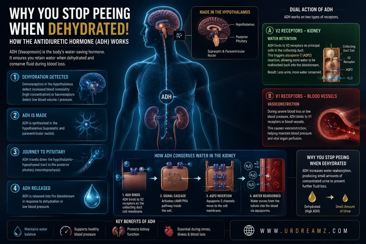
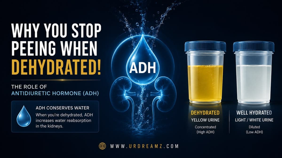
Have you ever noticed that when you haven't had enough water, your urine becomes darker, more concentrated, and far less frequent — and that after a big glass of water, everything reverses and your urine turns almost clear again? This is not a coincidence, and it is not your kidneys simply "running low." It is one specific hormone, manufactured deep inside your brain, actively deciding — moment to moment — exactly how much water your body is allowed to lose. That hormone is called Antidiuretic Hormone (ADH), also known as Vasopressin. This guide walks through its entire journey: where it is built, how it travels, how your brain actually detects dehydration in the first place, and the precise molecular chain of events inside your kidney that decides whether your urine comes out concentrated and orange, or diluted and clear.
1. What Is ADH, and Why Does Its Name Matter?
Antidiuretic Hormone (ADH) is found not only in humans but across essentially the entire animal kingdom, making it one of the more evolutionarily ancient and consistently conserved hormones in biology. Its name itself is a direct clue to exactly what it does. The word "diuresis" refers to water loss — specifically, the process of losing water through urine. The prefix "anti" simply means "against" or "preventing." Put the two together, and the name translates almost literally into plain English: this is the hormone that prevents water loss. Whenever the body senses that its water levels are running low, ADH is released specifically to stop further water from being lost through urine, conserving whatever water remains in the system.
This straightforward etymology is worth sitting with for a moment, because it captures the entire purpose of everything that follows in this guide. Every downstream mechanism described below — the specific brain nuclei that manufacture this hormone, the receptors that detect the need for it, and the elaborate cellular machinery inside the kidney that responds to it — exists in service of this one simple goal: stopping water from leaving the body when it is needed most. Understanding this core purpose up front makes every subsequent detail feel like a natural extension of a single, coherent idea, rather than a disconnected list of biological facts to memorize.
2. Where ADH Is Actually Made: Two Specific Brain Nuclei
ADH is manufactured directly within the hypothalamus, the small, almond-sized structure sitting deep within the brain that serves as the master regulatory center for a wide range of hormonal systems throughout the body. Within the hypothalamus, ADH production is not spread out diffusely — it is concentrated in two specific, named clusters of specialized neurons: the Supraoptic Nucleus and the Paraventricular Nucleus.
These two nuclei represent dense, tightly organized populations of specialized neurons whose entire job is to manufacture ADH (and, in the case of the paraventricular nucleus, also oxytocin) and then transport it onward for eventual release. The supraoptic nucleus sits, as its name suggests, just above the optic chiasm — the point where the two optic nerves from each eye partially cross paths — while the paraventricular nucleus sits closer to the third ventricle, one of the brain's fluid-filled cavities. Despite their slightly different locations within the hypothalamus, both nuclei contribute meaningfully to the body's total ADH output, working in a coordinated, complementary fashion rather than one substituting for the other.
3. The Journey Down: The Hypothalamic-Hypophyseal Tract
Once ADH is manufactured within these two hypothalamic nuclei, it does not simply diffuse randomly through brain tissue to reach its next destination. Instead, it travels along a specific, dedicated anatomical pathway called the hypothalamic-hypophyseal tract — essentially a bundle of long nerve fibers extending downward from the hypothalamus, through the thin connecting stalk, directly into the posterior lobe of the pituitary gland, where it is stored until needed.
This transport route is worth appreciating as a genuine biological engineering solution. The neurons whose cell bodies sit within the supraoptic and paraventricular nuclei have unusually long axons — the long, thin extensions of nerve cells that normally carry electrical signals — but in this specific case, these axons are also functioning as physical delivery conduits, carrying manufactured hormone molecules packaged in small vesicles down their entire length to the axon terminals sitting within the posterior pituitary. This is fundamentally different from how most of the body's other hormone-producing glands work, where the manufacturing site and the release site are typically the very same location.
4. Why ADH Is Also Called Vasopressin, AVP, and Arginine Vasopressin
ADH is frequently referred to by several alternative names in medical literature and clinical practice: Vasopressin, or more formally, AVP (Arginine Vasopressin). Understanding why requires looking directly at the hormone's molecular structure. ADH is a small peptide hormone, meaning it is built from a short chain of amino acids strung together — specifically, a chain of nine amino acids.
The specific identity of the amino acid occupying position eight in this nine-amino-acid chain is what gives the hormone its more formal name. In humans, the amino acid sitting at this eighth position is called arginine — hence "Arginine Vasopressin," or AVP for short. Interestingly, this is not universal across the animal kingdom: in pigs specifically, the amino acid occupying that same eighth position is a different amino acid called lysine instead, producing a molecule sometimes called lysine vasopressin. This single amino acid substitution represents essentially the only structural difference between the human and porcine versions of this otherwise nearly identical hormone — a small but genuinely fascinating detail illustrating how evolution can fine-tune an ancient, highly conserved hormone slightly differently across different species while preserving its overall function almost entirely intact.
5. From Preprohormone to Prohormone to Final ADH
ADH does not appear instantly in its final, active form the moment it is manufactured — it goes through a multi-step processing sequence first. Initially, the hypothalamic neurons produce a larger precursor molecule called a preprohormone. This preprohormone then undergoes an initial cutting or cleavage step, trimming it down into an intermediate form called a prohormone.
From this prohormone stage, further chemical modification takes place as the molecule travels down the long axon toward the posterior pituitary, ultimately producing the final, biologically active ADH molecule by the time it reaches its storage destination. This staged processing approach — building a larger, initially inactive precursor and then progressively refining it into its final functional form — is a common strategy used throughout the body for manufacturing many different peptide hormones, since it allows for additional layers of quality control and regulation at each processing step along the way, rather than risking the release of an incompletely formed, potentially non-functional hormone directly into circulation.
6. Neurophysin II: The Escort Protein
As ADH makes this journey from its site of manufacture down to its storage site in the posterior pituitary, it does not travel alone. It is bound to and transported alongside a specific carrier protein called Neurophysin II, which physically escorts the hormone along its journey and helps ensure it is properly packaged and delivered to its intended storage location within the posterior lobe.
Neurophysin II is produced from the very same larger preprohormone molecule that eventually yields ADH itself — both the final hormone and its escort protein originate from a single shared precursor, which is then cleaved into its separate functional components during the processing sequence described above. Once ADH and Neurophysin II arrive together at the posterior pituitary, they are stored together within small storage vesicles, remaining held in reserve until a signal arrives instructing the system to release the hormone into general circulation.
7. How the Hypothalamus Actually Knows You're Dehydrated
One of the most genuinely fascinating aspects of this entire system is the question of how the hypothalamus actually knows, in the first place, that the body needs more ADH released. The hypothalamus does not simply guess or operate on a fixed schedule — it continuously monitors specific, measurable signals arriving from throughout the body, using dedicated sensory structures called receptors, each tuned to detect a particular type of stimulus: temperature-sensing receptors, chemical-sensing receptors, and, most relevant to this guide, receptors specifically tuned to detect the body's water and blood pressure status.
This constant, ongoing monitoring is precisely what allows the hypothalamus to determine, moment to moment, exactly what the body currently needs — when to trigger a hormone release, and, just as importantly, when to hold back and stop signaling once a given need has already been addressed. Two specific detection systems are directly responsible for triggering ADH release, and each one deserves a detailed look on its own.
8. Osmoreceptors: Measuring Salt Concentration in Real Time
The first, and generally more sensitive, detection system involves specialized cells within the hypothalamus called osmoreceptors. These cells do not directly measure "how much water" is present in the body in absolute terms — instead, they measure something more precise and more immediately actionable: the concentration of dissolved salts and other particles within the surrounding blood, a property referred to in physiology as osmolality.
When the body loses water — through sweating, inadequate fluid intake, or any other route — the total volume of water in the blood decreases, while the amount of dissolved salt remains essentially the same. The mathematical consequence of this is straightforward: with less water diluting the same amount of salt, the blood's overall salt concentration, its osmolality, rises. Osmoreceptors are specifically built to detect this rise in osmolality with remarkable sensitivity, capable of registering even very small percentage increases in blood concentration.
9. Why Shrinking Cells Trigger the Alarm
The actual physical mechanism by which osmoreceptors detect rising blood osmolality is genuinely elegant: as the surrounding blood becomes more concentrated with salt relative to the fluid inside the osmoreceptor cells themselves, water is drawn out of these cells and into the now more concentrated surrounding blood, through the basic physical process of osmosis. This causes the osmoreceptor cells to physically shrink in size.
This shrinking is not merely incidental — it is the literal trigger mechanism itself. As these specialized cells shrink, this physical change directly activates electrical signaling within them, which in turn sends a direct signal to the nearby ADH-producing neurons within the hypothalamus, effectively communicating: "the surrounding fluid environment has become more concentrated — water is running low — release ADH." Through this cell-shrinkage-based mechanism, the hypothalamus is able to make its determination about the body's hydration status, and set the entire ADH release cascade into motion, within seconds of a meaningful change in blood osmolality occurring.
(High Osmolality)
Posterior Pituitary
Concentrated Urine
(Atria, Carotid, Aorta)
Posterior Pituitary
Smooth Muscle (V1)
BP Restored
10. Baroreceptors: The Second Detection System
Alongside osmoreceptors, the body relies on a second, complementary detection system built around specialized structures called baroreceptors. While osmoreceptors are specifically tuned to detect blood concentration (osmolality), baroreceptors are tuned to detect something different but closely related: blood pressure itself.
This second detection pathway matters because significant water or blood loss does not only affect blood concentration — it also directly reduces the total volume of fluid circulating through the cardiovascular system, which in turn causes blood pressure to drop. Baroreceptors provide the hypothalamus with a second, independent line of evidence that something is wrong with the body's fluid status, complementing and reinforcing the osmolality-based signal coming from the osmoreceptors.
11. Where Baroreceptors Actually Sit
Baroreceptors are located at several key points throughout the cardiovascular system: within the atria, the two upper receiving chambers of the heart; within the carotid arteries, the major blood vessels running up either side of the neck supplying blood to the brain; and within the aorta, the single largest blood vessel in the entire body, emerging directly from the heart and carrying oxygenated blood outward to the rest of the circulatory system.
Baroreceptors positioned at each of these strategic locations continuously monitor the pressure and stretch of the surrounding blood vessel walls. When blood volume drops — whether from dehydration, blood loss, or any other cause — blood pressure falls correspondingly, and the vessel walls stretch less than they normally would with each heartbeat. Baroreceptors detect this reduced stretch and respond by sending nerve signals to the hypothalamus, effectively communicating: "blood pressure is low — the body needs more water retained — release ADH."
12. Two Different Alarms, One Shared Response
It is worth appreciating that osmoreceptors and baroreceptors represent two genuinely independent detection systems, monitoring two different physiological variables — blood concentration versus blood pressure — yet both ultimately converge on the exact same response: triggering ADH release from the hypothalamus. This dual-detection design provides the body with a kind of built-in redundancy and cross-checking, ensuring that ADH release can be triggered appropriately whether the underlying problem is primarily one of water concentration (as in ordinary dehydration from inadequate fluid intake) or primarily one of volume loss (as in significant blood loss from injury), even though these two scenarios represent meaningfully different underlying physiological problems.
13. The Journey to the Kidney: Meet the Nephron
Once the hypothalamus has made its determination and triggered ADH release from the posterior pituitary, the hormone travels through the bloodstream directly to the kidneys. Each kidney contains approximately one million microscopic functional units called nephrons — meaning the two kidneys together contain roughly two million of these individual filtering-and-processing units, each one capable of independently filtering blood and producing urine.
A single nephron represents an extraordinarily sophisticated piece of biological engineering, comprising several distinct structural segments, each performing its own specific function in the overall process of filtering blood and producing urine. Understanding this structure is essential to understanding exactly where and how ADH ultimately exerts its water-conserving effect.
14. Following the Filtrate Through the Nephron
Blood filtration begins at a structure called the glomerulus, a tight tangle of tiny blood vessels enclosed within a cup-shaped structure called Bowman's capsule. Here, blood pressure forces water, salts, and small dissolved molecules out of the blood and into the nephron's tubule system, while larger components like blood cells and proteins remain behind in the bloodstream. This filtered fluid, now called filtrate, then travels sequentially through the proximal convoluted tubule, down through a long, hairpin-shaped structure called the loop of Henle, and then up through the distal convoluted tubule, before finally reaching the structure most relevant to this entire guide: the collecting duct.
Along most of this journey, a substantial amount of water and useful solutes are already reabsorbed back into the bloodstream through various mechanisms independent of ADH. However, the truly critical, adjustable decision point regarding exactly how much additional water gets reabsorbed — or allowed to pass onward and be excreted as urine — happens specifically at the final structure in this sequence: the collecting duct.
15. Arriving at the Collecting Duct
The collecting duct is the final structure through which filtrate passes before it becomes what we ultimately recognize and pass as urine. Anatomically, it has a distinctive pinkish appearance under microscopic examination, and it serves as the specific site where ADH exerts its primary, defining effect on the body's water balance.
It is worth emphasizing just how much regulatory power is concentrated at this single anatomical checkpoint. Rather than water reabsorption being fixed and unchangeable throughout the nephron, the collecting duct functions as a genuinely adjustable final gate — permeable to water when ADH is present and actively signaling, and comparatively far less permeable to water when ADH levels are low. This single adjustable checkpoint is what gives the body its remarkable, moment-to-moment flexibility in deciding exactly how much water to conserve versus excrete, based on current physiological need.
16. The V2 Receptor: ADH's Docking Station
Embedded within the cell membranes lining the collecting duct are specialized docking sites called V2 receptors, specifically designed to recognize and bind circulating ADH molecules. When ADH arrives at the collecting duct via the bloodstream, it binds directly to these V2 receptors, initiating the entire downstream cellular cascade responsible for increasing water reabsorption.
This receptor-based system represents a classic example of hormonal signal specificity — V2 receptors are specifically shaped to recognize and respond to ADH and essentially nothing else circulating in the bloodstream, ensuring that this particular water-conservation mechanism activates only in direct response to genuine ADH signaling, rather than being accidentally triggered by unrelated circulating substances.
17. Inside the Cell: Adenylate Cyclase and the cAMP Surge
Once ADH successfully binds to a V2 receptor on the outer surface of a collecting duct cell, this binding event triggers activation of an enzyme located within the cell membrane called Adenylate Cyclase (AC). Once activated, this enzyme rapidly increases the concentration of a specific internal signaling molecule called cAMP (cyclic Adenosine Monophosphate) throughout the interior of the cell.
This cAMP surge functions as what biologists often refer to as a "second messenger" — the original ADH signal arrived and bound at the outer cell surface, but cAMP is what actually carries and amplifies that signal onward into the cell's interior, translating an external hormonal message into a specific internal biochemical instruction the cell's own machinery can directly act upon.
18. PKA Gets Activated
As cAMP levels rise within the collecting duct cell, this rising concentration activates yet another protein, called PKA (Protein Kinase A). Once activated, PKA continues driving the signaling cascade forward, ultimately triggering the specific structural change within the cell that will directly determine how much water actually gets reabsorbed.
This multi-step signaling relay — ADH binding to V2, triggering adenylate cyclase, raising cAMP, activating PKA — might initially seem like an unnecessarily complicated chain of intermediate steps to accomplish a single outcome. In reality, this kind of multi-step cascade allows for substantial signal amplification along the way: a single ADH molecule binding to a single V2 receptor can, through this cascading activation sequence, ultimately influence the behavior of a very large number of downstream target molecules within that same cell, allowing a relatively small amount of circulating hormone to produce a meaningfully large physiological effect.
19. Aquaporin-2: The Water Doors That Change Everything
The final, defining step in this entire cascade involves a specific protein called Aquaporin-2 — literally, a water channel or "water door" embedded within small internal storage vesicles inside the collecting duct cell. Once PKA becomes active, it triggers these Aquaporin-2-containing vesicles to move toward and fuse directly with the inward-facing surface of the cell membrane — the side facing into the collecting duct's internal tubule space.
Once this fusion occurs, the Aquaporin-2 water channels become embedded directly in that inward-facing cell membrane, creating genuine, functional water-permeable pathways where none previously existed. With these channels newly in place, water sitting within the collecting duct tubule — water that would otherwise have simply continued onward and been excreted as urine — is now able to pass directly through these channels, out of the tubule, through the cell, and back into the surrounding blood supply, effectively being reclaimed by the body rather than lost.
20. Why Dehydrated Urine Turns Orange and Concentrated
This entire mechanism directly explains one of the most familiar, everyday signs of dehydration: dark, concentrated, orange-tinted urine. When the body is dehydrated, ADH levels rise significantly, driving large numbers of Aquaporin-2 channels into the collecting duct's cell membrane, pulling a substantial amount of water back out of the forming urine and into the bloodstream. What remains behind in the collecting duct — and is ultimately excreted as urine — is the same total amount of waste product (urea, salts, and other metabolic byproducts) now concentrated into a much smaller remaining volume of water, producing the characteristic darker, more concentrated, orange-toned appearance.
21. Why Well-Hydrated Urine Turns Clear and Dilute
The exact opposite process occurs when the body has plenty of water available. In this well-hydrated state, ADH release drops significantly, since the osmoreceptors and baroreceptors are no longer detecting any meaningful water deficit. With lower ADH levels, fewer Aquaporin-2 channels are inserted into the collecting duct membrane, meaning far less water gets pulled back out of the forming urine. The result is urine that remains far more dilute — pale yellow to nearly clear — since the same waste products are now dispersed throughout a much larger remaining volume of water that was never reclaimed by the body.
22. The Emergency Scenario: Severe Blood Loss
Beyond ordinary, everyday dehydration, ADH plays an even more dramatic role in a genuine medical emergency: significant blood loss, such as might occur following a serious accident or traumatic injury. In this scenario, blood pressure can drop rapidly and severely as circulating blood volume decreases, triggering an intense baroreceptor-driven signal to the hypothalamus, resulting in dramatically elevated ADH release — far beyond the levels typically seen in ordinary, everyday dehydration.
23. Enter the V1 Receptor: A Completely Different Job
At these emergency-level concentrations, ADH does not act solely through the V2 receptors and collecting duct mechanism described earlier — it also begins binding to a second, entirely distinct type of receptor called the V1 receptor. Unlike V2 receptors, which are located specifically in the kidney's collecting duct, V1 receptors are located within the smooth muscle lining the walls of blood vessels throughout the body.
24. Vasoconstriction: Squeezing the Blood Vessels
When ADH binds to these V1 receptors on blood vessel smooth muscle, it triggers those muscles to contract, narrowing the internal diameter of the affected blood vessels — a process called vasoconstriction. This vessel-narrowing effect directly helps counteract the drop in blood pressure caused by significant blood loss, since narrower blood vessels require less total blood volume to maintain adequate pressure throughout the circulatory system, similar to how a narrower hose can maintain stronger water pressure than a wider one carrying the same flow rate.
25. Why This Emergency Response Can Be Lifesaving
This V1-receptor-driven vasoconstriction response is not a minor, incidental side effect — it can be genuinely lifesaving in the setting of severe blood loss. If blood pressure is allowed to fall too far without this compensatory mechanism, blood flow to the brain becomes inadequate, risking cardiac arrest, stroke, or permanent brain injury from oxygen deprivation. By actively narrowing blood vessels throughout the body during a genuine volume crisis, ADH's V1-mediated action helps preserve enough blood pressure to maintain adequate perfusion to the brain and heart during the critical window before medical treatment, such as fluid or blood replacement, can be administered.
26. What Happens When ADH Goes Wrong: Diabetes Insipidus and SIADH
Given how central ADH is to water balance, it should come as no surprise that malfunctions in this system produce genuine, recognizable medical conditions. When the body produces too little ADH, or the kidneys fail to respond to it properly, the resulting condition is called Diabetes Insipidus — characterized by an inability to retain water, leading to the passage of unusually large volumes of very dilute, pale urine, alongside persistent, often extreme thirst. At the opposite extreme, when the body produces and releases too much ADH, the resulting condition is called SIADH (Syndrome of Inappropriate Antidiuretic Hormone secretion), in which the body retains far too much water, diluting the blood's sodium concentration to potentially dangerous levels and increasing the risk of swelling and other complications.
| Condition | ADH Level | Key Effect |
|---|---|---|
| Diabetes Insipidus | Too low (or kidney doesn't respond) | Excess water loss, large volumes of dilute urine, extreme thirst |
| SIADH | Too high | Excess water retention, diluted blood sodium, risk of dangerous swelling |
27. Putting the Entire System Together
Bringing every step of this guide together into a single continuous story: the hypothalamus manufactures ADH within the supraoptic and paraventricular nuclei, transports it down the hypothalamic-hypophyseal tract for storage in the posterior pituitary, and continuously monitors the body's hydration and blood pressure status through osmoreceptors and baroreceptors. When either system detects a genuine water or volume deficit, ADH is released into the bloodstream, traveling to the kidney's collecting ducts, where it binds V2 receptors, triggers a cAMP-and-PKA signaling cascade, and ultimately inserts Aquaporin-2 water channels that pull water back into the body — producing the darker, more concentrated urine everyone recognizes as a sign of dehydration. In genuine emergencies involving significant blood loss, this same hormone additionally activates V1 receptors on blood vessels, directly helping preserve blood pressure and, in doing so, potentially saving a life. From a single hormone's name meaning simply "against water loss," this entire elegant, multi-layered system unfolds.
28. Frequently Asked Questions
- Q1: What does ADH stand for?
- ADH stands for Antidiuretic Hormone, also known as Vasopressin or Arginine Vasopressin (AVP).
- Q2: Where is ADH made in the body?
- ADH is made in the hypothalamus, specifically within the Supraoptic Nucleus and Paraventricular Nucleus, and is then stored in the posterior pituitary gland.
- Q3: Why is ADH also called Arginine Vasopressin?
- Because in humans, the amino acid at position eight of its nine-amino-acid chain is arginine; in pigs, this same position is occupied by lysine instead.
- Q4: What are osmoreceptors?
- Osmoreceptors are specialized cells in the hypothalamus that detect blood concentration (osmolality) by shrinking when blood becomes more concentrated, triggering ADH release.
- Q5: What are baroreceptors, and where are they located?
- Baroreceptors detect blood pressure and are located in the atria of the heart, the carotid arteries, and the aorta; low blood pressure triggers them to signal for ADH release.
- Q6: What is a nephron?
- A nephron is the microscopic functional unit of the kidney responsible for filtering blood and producing urine; each kidney contains approximately one million nephrons.
- Q7: Where in the kidney does ADH act?
- ADH acts specifically on the collecting duct, the final segment of the nephron, where it increases water reabsorption back into the blood.
- Q8: What is the V2 receptor?
- The V2 receptor is a docking site on collecting duct cells that binds ADH, triggering a signaling cascade involving cAMP and PKA that leads to increased water reabsorption.
- Q9: What is Aquaporin-2?
- Aquaporin-2 is a water channel protein that, once inserted into the collecting duct cell membrane in response to ADH, allows water to be reabsorbed from urine back into the blood.
- Q10: Why does dehydration cause dark, orange urine?
- Higher ADH levels during dehydration cause more water to be reabsorbed via Aquaporin-2 channels, concentrating the same waste products into a smaller volume of remaining urine.
- Q11: What is the V1 receptor, and how is it different from V2?
- V1 receptors are located on blood vessel smooth muscle and, at high ADH levels, trigger vasoconstriction to help maintain blood pressure, unlike V2 receptors which act on the kidney.
- Q12: How does ADH help during severe blood loss?
- Severe blood loss triggers very high ADH release, which activates V1 receptors to constrict blood vessels, helping preserve blood pressure and blood flow to vital organs like the brain.
- Q13: What is Diabetes Insipidus?
- Diabetes Insipidus is a condition caused by too little ADH (or kidney unresponsiveness to it), resulting in excessive water loss, large volumes of dilute urine, and extreme thirst.
- Q14: What is SIADH?
- SIADH (Syndrome of Inappropriate Antidiuretic Hormone secretion) is a condition caused by excessive ADH release, leading to excess water retention and dangerously diluted blood sodium.
- Q15: Why does urine become clear when well-hydrated?
- With adequate hydration, ADH levels drop, fewer Aquaporin-2 channels are active, less water is reabsorbed, and the same waste products are dispersed in a larger volume of water, producing pale, dilute urine.
29. Conclusion
The simple act of noticing that your urine has become darker after a long day without enough water is, in reality, the visible endpoint of an extraordinarily intricate hormonal system — one that begins with two specific clusters of neurons deep in the hypothalamus, travels down a dedicated nerve pathway to the pituitary gland, is triggered by two independent detection systems monitoring blood concentration and blood pressure, and ultimately reaches into the microscopic collecting ducts of two million individual kidney nephrons to physically insert water channels that reclaim water your body would otherwise lose. In moments of genuine crisis, this same hormone shifts into an entirely different mode, tightening blood vessels throughout the body to help preserve blood pressure and keep the brain and heart supplied with blood. From a name that simply means "against water loss," ADH reveals itself to be one of the most elegantly engineered, life-preserving systems in all of human physiology.