Spermatogenesis & Testicular Temperature: The Complete Molecular Physiology of How Sperm Are Actually Made
Welcome back to Lecture 2 of our Human Reproduction series. Our first lecture examined a fully mature sperm cell's structure in complete anatomical detail. This lecture rewinds the clock even further, to the very beginning of that cell's existence — the process called spermatogenesis, through which the testes continuously manufacture new sperm cells from a small, self-renewing population of stem cells. This is a genuinely remarkable production line: every single sperm cell takes exactly 74 days to build, runs under the tight hormonal control of a chain of command stretching from the brain all the way down to two distinct testicular cell types, and depends entirely on a temperature requirement most people never think twice about — the testes must stay 2 to 4°C cooler than the rest of the body, or the entire process fails.
In this lecture, we'll trace the complete journey of a developing sperm cell — from spermatogonium through primary spermatocyte, secondary spermatocyte, and spermatid, to a fully formed spermatozoon — and examine the essential supporting roles played by Sertoli cells, Leydig cells, FSH, LH, and testosterone throughout that entire journey. We'll then turn to an equally fascinating engineering problem: how the body actually keeps the testes cooler than everywhere else, through three distinct, coordinated anatomical systems — the pampiniform plexus, the cremaster muscle, and the dartos muscle.
Quick Answer
Last updated: September 2026
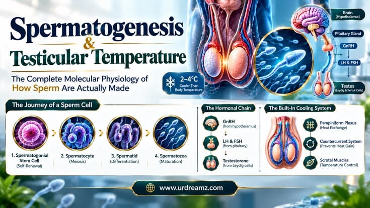
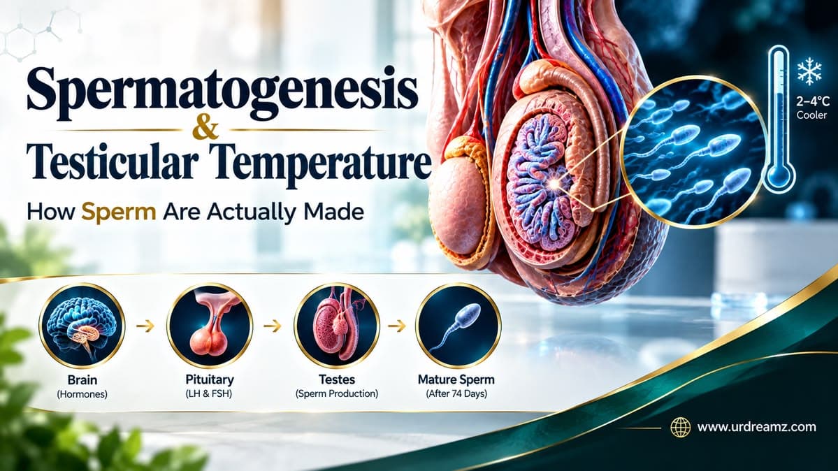
Spermatogenesis is the roughly 74-day process by which the testes produce sperm, progressing through four cell stages: spermatogonia (diploid stem cells) divide by mitosis and enter meiosis as primary spermatocytes, which complete meiosis I to form secondary spermatocytes, which complete meiosis II to form haploid spermatids, which then undergo spermiogenesis to become mature spermatozoa. This process is supported by two key testicular cell types: Sertoli cells, which nourish and physically support developing sperm cells and respond to follicle-stimulating hormone (FSH), and Leydig cells, located between the seminiferous tubules, which produce testosterone in response to luteinizing hormone (LH). Both FSH and LH are released by the pituitary gland in response to gonadotropin-releasing hormone (GnRH) from the hypothalamus, forming the hypothalamic-pituitary-gonadal (HPG) axis. Because sperm development is highly temperature-sensitive, the testes must be kept 2 to 4°C cooler than core body temperature, a task accomplished by three coordinated systems: the pampiniform plexus (a network of veins that cools incoming arterial blood through countercurrent heat exchange), the cremaster muscle (which raises or lowers the testes relative to the body based on temperature), and the dartos muscle (which contracts or relaxes the scrotal skin to adjust surface area for heat loss).
1. Welcome Back: Where Sperm Actually Begin
Every mature sperm cell begins its existence deep inside the testis, within a tightly coiled structure called the seminiferous tubule. This lecture traces that entire developmental journey from beginning to end — a process that, remarkably, never fully stops throughout a man's reproductive life, continuously generating new sperm cells from a small, dedicated population of stem cells that themselves are never entirely used up.
2. The Testis From the Inside: Seminiferous Tubules
Each testis is packed with hundreds of tightly coiled seminiferous tubules, and it's within the walls of these tubules that the entire spermatogenesis process, covered step by step throughout this lecture, actually takes place. Developing sperm cells progress through their various stages while moving from the outer edge of the tubule wall inward, toward the central open space (the lumen), where fully formed sperm are eventually released to begin their onward journey.
3. Spermatogonia: The Stem Cells That Never Run Out
At the very outer edge of each seminiferous tubule sit spermatogonia — diploid stem cells, carrying the full 46-chromosome complement, that serve as the entire foundation of ongoing sperm production. These cells are genuinely special because they maintain their own numbers indefinitely: rather than being gradually depleted over a man's lifetime, spermatogonia continuously divide to produce both new spermatogonia (maintaining the stem cell reserve) and cells committed to entering the developmental pathway traced throughout the rest of this lecture.
4. Mitosis First: Keeping the Stem Cell Pool Alive
Before any cell commits to becoming a sperm, spermatogonia first divide by ordinary mitosis, the same basic cell division process used throughout most of the body for routine growth and tissue maintenance. This mitotic division serves two simultaneous purposes: it replenishes the spermatogonial stem cell population, ensuring the supply never runs out, while also producing daughter cells that will go on to commit fully to the specialized developmental pathway that follows.
5. Primary Spermatocytes: Entering Meiosis I
Once a spermatogonium commits to development, it grows and transforms into a primary spermatocyte — still diploid at this stage, carrying the full 46 chromosomes, but now entering meiosis, the specialized cell division process responsible for ultimately halving the chromosome number to produce genetically unique gametes. The primary spermatocyte undergoes meiosis I specifically, during which genetic recombination occurs (contributing to genetic diversity, as covered in our earlier lecture on sperm-ovum fusion) and the cell divides into two daughter cells.
6. Secondary Spermatocytes: The Brief Middle Stage
The two daughter cells produced by meiosis I are called secondary spermatocytes. Each now carries only 23 chromosomes — technically haploid, though each chromosome still consists of duplicated sister chromatids at this stage. Secondary spermatocytes are notably short-lived, existing only briefly before rapidly proceeding into the second meiotic division covered in the next section.
7. Spermatids: Meiosis II Completes the Chromosome Halving
Secondary spermatocytes undergo meiosis II, a division process that separates the duplicated sister chromatids from one another, producing spermatids — genuinely haploid cells, each carrying a single, complete set of 23 chromosomes, precisely the chromosome number required for the fertilization process covered in detail in our earlier lecture on sperm-ovum fusion. At this stage, spermatids are still round, relatively unspecialized cells, structurally quite different from the streamlined, tailed sperm cell described in our first lecture.
(Diploid, 46 chr.)
(Meiosis I)
(Haploid, 23 chr.)
(Meiosis II Complete)
(Spermiogenesis)
8. Spermiogenesis: From Round Cell to Swimming Cell
The final stage of this journey is spermiogenesis — the dramatic structural transformation through which a round, unremarkable spermatid becomes a fully formed, functional spermatozoon. As covered in detail in our first lecture on sperm anatomy, this transformation includes the development of the acrosome, the compaction of DNA using protamines in place of histones, the shedding of excess cytoplasm (later phagocytosed by Sertoli cells, covered in the next section), and the growth of the characteristic tail structure responsible for swimming motion. This is the single most visually dramatic stage of the entire process, converting a genuinely unremarkable-looking cell into the highly specialized structure this series has examined in such detail.
9. The Complete 74-Day Timeline
Taken together, the entire spermatogenesis process — from a committed spermatogonium through to a fully released, mature spermatozoon — takes approximately 74 days to complete in humans. This is a remarkably consistent, tightly regulated biological timeline, and it carries genuine practical significance: any factor affecting testicular health at a given point in time — illness, heat exposure, certain medications — may not show its effect on semen quality until roughly two and a half months later, once the affected cohort of developing sperm cells finally completes this entire journey and is released.
| Stage | Approximate Duration |
|---|---|
| Spermatogonium to Primary Spermatocyte | Ongoing mitotic renewal phase |
| Meiosis I (Primary → Secondary Spermatocyte) | Several weeks |
| Meiosis II (Secondary Spermatocyte → Spermatid) | Brief, rapid transition |
| Spermiogenesis (Spermatid → Spermatozoon) | Several weeks |
| Total Process | ~74 days |
10. Sertoli Cells: The Nurse Cells That Run the Nursery
None of the developmental journey covered so far happens in isolation — it unfolds under the close structural and nutritional support of Sertoli cells, large support cells embedded within the seminiferous tubule wall, extending from the outer edge all the way to the central lumen. Sertoli cells are sometimes called "nurse cells" precisely because of the role they play: physically supporting developing sperm cells at every stage, supplying them with nutrients, and, as covered in our first lecture, absorbing and clearing away excess cytoplasm shed during spermiogenesis through phagocytosis.
11. The Blood-Testis Barrier: A Protective Wall
Sertoli cells also form tight junctions with one another, creating a genuinely important structure called the blood-testis barrier. This barrier physically separates the outer portion of the seminiferous tubule from the inner, more advanced developmental stages, creating an immunologically privileged environment shielded from the body's own immune system. This matters because developing sperm cells, particularly once haploid, express surface proteins the immune system could potentially recognize as foreign, since they emerge only after puberty, well after the immune system has already established what counts as "self." The blood-testis barrier prevents this kind of immune reaction from ever developing in the first place.
12. Leydig Cells: The Testosterone Factory
Situated in the interstitial tissue between the seminiferous tubules, rather than within their walls, sit Leydig cells — the testes' dedicated testosterone-producing cells. Unlike Sertoli cells, which directly support developing sperm structurally, Leydig cells' primary role is hormonal: producing testosterone, which, as covered in the sections ahead, plays an essential role both locally within the testis and throughout the rest of the body.
| Feature | Sertoli Cells | Leydig Cells |
|---|---|---|
| Location | Within seminiferous tubule walls | Interstitial tissue, between tubules |
| Primary Role | Structural/nutritional support for developing sperm | Testosterone production |
| Responds To | FSH (follicle-stimulating hormone) | LH (luteinizing hormone) |
| Additional Function | Forms blood-testis barrier; secretes inhibin | Supports local testosterone concentration needed for spermatogenesis |
13. The HPG Axis: Hypothalamus, Pituitary, and Gonads
The entire hormonal system driving spermatogenesis operates through a coordinated chain of command called the hypothalamic-pituitary-gonadal (HPG) axis, involving three distinct anatomical levels working in careful sequence: the hypothalamus in the brain, the pituitary gland just below it, and the testes (gonads) themselves.
14. GnRH: The Signal That Starts It All
The hypothalamus releases gonadotropin-releasing hormone (GnRH) in a pulsatile pattern, traveling a short distance to the nearby pituitary gland. This release isn't constant or steady — it occurs in rhythmic pulses, and this specific pulsatile pattern is itself biologically important, since continuous, non-pulsatile GnRH exposure actually suppresses, rather than stimulates, the downstream hormonal response covered in the following sections.
15. FSH: Talking Directly to Sertoli Cells
In response to GnRH stimulation, the pituitary gland releases follicle-stimulating hormone (FSH) into the bloodstream, which travels to the testes and binds specifically to receptors on Sertoli cells, discussed earlier in this lecture. This FSH signal is essential for supporting Sertoli cell function, including their nutritional support of developing sperm cells and their role in maintaining the blood-testis barrier — meaning FSH's influence on spermatogenesis is indirect, working through Sertoli cells rather than acting on developing sperm cells themselves.
16. LH: Talking Directly to Leydig Cells
Alongside FSH, the pituitary also releases luteinizing hormone (LH), which travels to the testes and binds specifically to receptors on Leydig cells, stimulating them to produce testosterone. This is the specific hormonal signal responsible for driving the testosterone production discussed earlier in this lecture, completing the second major branch of the HPG axis's downstream signaling.
(GnRH)
(FSH + LH)
LH → Leydig Cells
+ Testosterone Production
17. Testosterone's Dual Role: Local and Systemic
Testosterone produced by Leydig cells plays two genuinely distinct roles simultaneously. Locally, within the testis itself, high concentrations of testosterone are essential for supporting the spermatogenesis process covered throughout the earlier sections of this lecture — concentrations considerably higher than what circulates in the bloodstream generally. Systemically, testosterone released into general circulation is responsible for the broader range of male secondary sexual characteristics and overall physiological effects typically associated with the hormone, extending well beyond the testis itself.
18. Inhibin and Negative Feedback: Keeping the System Balanced
The HPG axis doesn't operate as a one-way signal alone — it includes a feedback system that keeps hormone levels appropriately balanced. Sertoli cells secrete a hormone called inhibin, which travels back to the pituitary gland and specifically suppresses further FSH release, providing a direct feedback signal reflecting the current state of spermatogenesis. Testosterone itself also feeds back to suppress GnRH and LH release, at both the hypothalamus and pituitary level. Together, these feedback loops keep the entire hormonal system operating within an appropriate, tightly regulated range, rather than allowing hormone levels to rise or fall unchecked.
19. Why Temperature Matters So Much for Sperm
Having covered the complete cellular and hormonal story of spermatogenesis, this lecture now turns to a second, equally essential requirement: temperature. Spermatogenesis is a genuinely temperature-sensitive process, and the enzymes and cellular machinery involved in the various developmental stages covered earlier in this lecture function optimally only within a fairly narrow temperature range — one that happens to be cooler than the rest of the human body. This is precisely why the testes are positioned externally, within the scrotum, rather than internally alongside the rest of the reproductive organs.
20. The 2–4°C Rule: How Cool Is Cool Enough
Specifically, the testes must be maintained at a temperature roughly 2 to 4°C cooler than core body temperature for spermatogenesis to proceed normally. Even relatively modest, sustained increases above this target range can measurably impair sperm production and quality, which is precisely why the body invests in the three distinct, coordinated anatomical systems covered in the remaining sections of this lecture, specifically dedicated to maintaining this precise temperature difference around the clock.
21. The Pampiniform Plexus: A Living Heat Exchanger
The first of these three systems is the pampiniform plexus, a dense network of veins surrounding the testicular artery as it travels down toward the testis. This structure functions as a genuinely elegant biological heat exchanger: relatively warm arterial blood traveling downward toward the testis passes in close proximity to cooler venous blood traveling upward, away from the testis, and heat transfers directly from the warmer artery to the cooler surrounding veins before the arterial blood ever reaches the testis itself.
22. Countercurrent Exchange Explained Simply
This mechanism is called countercurrent heat exchange, and it works specifically because the two blood flows move in opposite directions, in close physical contact with one another. As warm arterial blood flows downward and cooler venous blood flows upward alongside it, heat continuously transfers from the warmer to the cooler stream along their entire shared length, meaning the arterial blood arriving at the testis has already been pre-cooled considerably compared to blood elsewhere in the body — a passive, continuously operating cooling system requiring no active energy expenditure beyond the blood flow itself.
Flows Downward
Venous Blood (Upward)
Artery to Vein
Reaches Testis
23. The Cremaster Muscle: An Elevator for the Testes
The second temperature-regulating system is the cremaster muscle, a thin band of skeletal muscle surrounding the spermatic cord and testis. This muscle can contract, pulling the testis upward, closer to the body, in cold conditions, where core body heat can help maintain adequate warmth — or relax, allowing the testis to descend further from the body in warm conditions, where greater distance helps maintain the necessary cooling. This muscle also underlies the well-known cremasteric reflex, a rapid, involuntary contraction triggered by touch or cold, that clinicians commonly test as part of a standard physical examination.
24. The Dartos Muscle: Wrinkling for Warmth
The third and final system is the dartos muscle, a layer of smooth muscle within the scrotal skin itself, distinct from the skeletal cremaster muscle discussed above. In cold conditions, the dartos muscle contracts, causing the scrotal skin to wrinkle and tighten, reducing its overall surface area and pulling it closer to the body to conserve heat. In warm conditions, it relaxes, allowing the scrotum to hang more loosely, increasing surface area and promoting heat loss to the surrounding environment.
| System | Type | Mechanism |
|---|---|---|
| Pampiniform Plexus | Vascular (passive heat exchange) | Cools arterial blood via countercurrent exchange with venous blood |
| Cremaster Muscle | Skeletal muscle | Raises or lowers testes relative to the body |
| Dartos Muscle | Smooth muscle (scrotal skin) | Wrinkles or relaxes scrotal skin to adjust surface area |
25. What Happens When This System Fails: Cryptorchidism and Varicocele
Understanding this system's normal operation also helps explain two related clinical conditions. Cryptorchidism occurs when one or both testes fail to descend into the scrotum during fetal development, remaining instead within the warmer abdominal cavity — a condition that, left uncorrected, can significantly impair future spermatogenesis precisely because the temperature requirement covered throughout this lecture is never actually met. Varicocele, by contrast, involves an abnormal enlargement of veins within the pampiniform plexus itself, which can disrupt the countercurrent heat exchange mechanism described earlier, allowing testicular temperature to rise above its optimal range and, in some cases, measurably affecting fertility.
26. Putting It All Together: A System of Systems
Stepping back from the individual mechanisms covered throughout this lecture, spermatogenesis emerges as a genuinely remarkable example of biological coordination: a precisely timed 74-day cellular developmental sequence, supported by two distinct specialized cell types, governed by a multi-level hormonal feedback system reaching all the way from the brain, and entirely dependent on a dedicated, three-part anatomical cooling system operating continuously in the background. None of these individual pieces — the cell division sequence, the Sertoli-Leydig cell partnership, the HPG axis, or the temperature regulation system — would be sufficient on its own; it's the coordinated interaction of all of them together that makes normal, ongoing sperm production possible.
27. Frequently Asked Questions
What are the main stages of spermatogenesis?
Spermatogonia develop into primary spermatocytes (meiosis I), then secondary spermatocytes, then spermatids (meiosis II), which undergo spermiogenesis to become mature spermatozoa.
How long does spermatogenesis take?
The complete process takes approximately 74 days in humans.
What is the difference between Sertoli cells and Leydig cells?
Sertoli cells provide structural and nutritional support to developing sperm and respond to FSH, while Leydig cells produce testosterone in response to LH.
What is the blood-testis barrier?
It is a barrier formed by tight junctions between Sertoli cells that protects developing sperm cells from the immune system.
What is the HPG axis?
The hypothalamic-pituitary-gonadal axis is the hormonal chain of command involving GnRH from the hypothalamus, FSH and LH from the pituitary, and testosterone from the testes.
Why must the testes be cooler than the rest of the body?
Spermatogenesis is temperature-sensitive and functions optimally about 2 to 4°C cooler than core body temperature.
What is the pampiniform plexus and how does it work?
It is a network of veins that cools arterial blood traveling to the testis through countercurrent heat exchange with cooler venous blood flowing in the opposite direction.
What is the difference between the cremaster and dartos muscles?
The cremaster is a skeletal muscle that raises or lowers the testes relative to the body, while the dartos is a smooth muscle in the scrotal skin that wrinkles or relaxes to adjust surface area for heat loss.
What is cryptorchidism?
It is a condition where one or both testes fail to descend into the scrotum, which can impair spermatogenesis due to the warmer abdominal temperature.
What is a varicocele?
It is an abnormal enlargement of veins in the pampiniform plexus that can disrupt testicular temperature regulation and affect fertility.