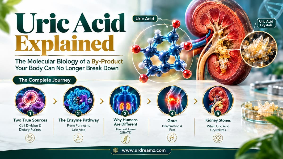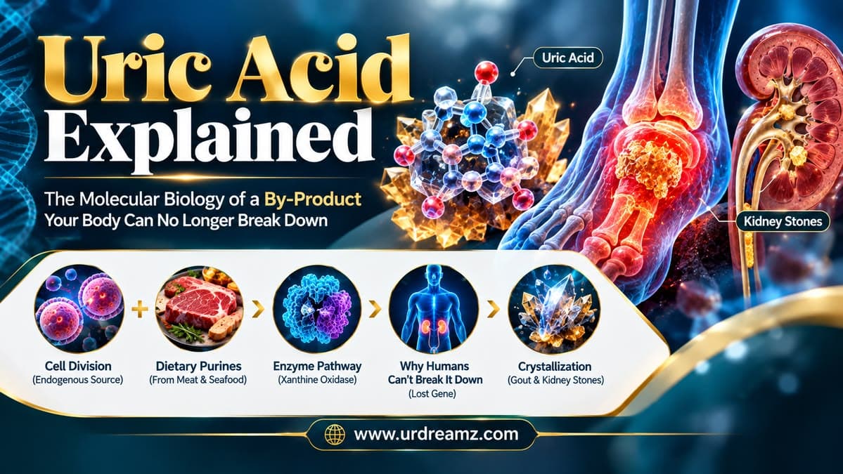
Uric Acid Explained: The Molecular Biology of a By-Product Your Body Can No Longer Break Down
Somewhere inside your body, right now, cells are dividing, dying, and being replaced — a completely normal, continuous process that has been running since before you were born. Every single time this happens, the DNA and RNA inside those cells get broken down, and a specific molecular by-product is left behind: uric acid. This isn't a mistake, a toxin, or something your body shouldn't be making — it's a completely normal, expected leftover of ordinary cellular turnover. The real problem isn't that uric acid exists. The real problem is that, for a very specific evolutionary reason we'll trace in detail in this article, the human body lost its ability to properly dissolve and dispose of it millions of years ago — a permanent biological inheritance every single person alive today still carries.
In this molecular-level guide, we'll trace uric acid all the way from its true origin — inside your own dividing cells — through the exact enzyme pathway responsible for creating it, examine the second, dietary source contributing to your total uric acid load, explain precisely why humans are uniquely vulnerable to this molecule in a way most animals simply aren't, and walk through exactly how it crystallizes into two genuinely painful conditions: kidney stones and gout.
Quick Answer
Last updated: September 2026
Uric acid is a natural waste product formed when purines — components of DNA and RNA — are broken down. Roughly 70% comes from your own body's routine cell turnover (endogenous production), while the remaining 30% comes from purine-rich foods like red meat and organ meats (exogenous/dietary sources). The enzyme xanthine oxidase converts purine breakdown products (hypoxanthine and xanthine) into uric acid, primarily in the liver. Unlike most animals, humans lack a functional uricase enzyme, which would normally convert uric acid into a far more water-soluble compound called allantoin — a gene humans lost through evolution millions of years ago. Because uric acid dissolves poorly in blood and urine, when levels rise too high, or urine becomes too acidic or concentrated, it can crystallize — forming kidney stones (accounting for roughly 10% of all kidney stone cases) or, when monosodium urate crystals deposit in joints (classically the big toe), triggering the intensely painful inflammatory condition known as gout. Management involves reducing purine-rich foods, staying well hydrated, and, when needed, medications like allopurinol or febuxostat that block uric acid production, or probenecid, which helps the kidneys excrete more of it.
1. Meet Uric Acid: The Body's Own Leftover
Uric acid is a naturally occurring chemical compound, produced whenever the body breaks down substances called purines. Far from being an abnormal toxin, it's a completely routine metabolic by-product — the kind of leftover molecule that appears whenever cellular material is broken down and processed, similar in concept to other waste products the body regularly generates and disposes of. What makes uric acid worth an entire dedicated discussion isn't its existence, but rather what happens when the body produces more of it than it can efficiently clear, a topic this article will trace all the way down to the molecular level.
2. Two Sources, One Molecule: Endogenous vs. Exogenous
Every molecule of uric acid circulating in your blood right now traces back to one of exactly two sources. Endogenous production — uric acid generated internally, by the body's own routine cellular processes — accounts for roughly 70% of the total. Exogenous production — uric acid derived from purine-containing foods and drinks consumed in the diet — accounts for the remaining 25 to 30%. Understanding this split matters enormously for managing uric acid levels, since it explains why dietary changes alone, while genuinely helpful, often cannot fully resolve high uric acid on their own — a meaningful majority of the total supply is being generated by the body itself, independent of what's on the plate.
*Commonly cited approximate split in metabolic and nephrology literature.
3. Where Endogenous Uric Acid Really Comes From: Cell Turnover
The body's own internal uric acid production is directly tied to one of the most fundamental processes in all of biology: cell division. Every time a cell in your body divides — and this is happening constantly, across nearly every tissue — its DNA and RNA must be processed, and old genetic material is broken down as part of that process. This breakdown releases specific molecular building blocks, and it's from these building blocks that the majority of your body's uric acid actually originates, entirely independent of anything you eat.
4. DNA's Building Blocks: Purines and Pyrimidines
DNA and RNA are built from smaller molecular units called nucleotides, and each nucleotide contains a nitrogen-containing base belonging to one of two chemical families: purines and pyrimidines. Uric acid specifically originates from the purine family — not from pyrimidines, which are broken down through an entirely separate metabolic pathway. The two purine bases relevant to this entire discussion are adenine and guanine, both found within the genetic material of essentially every cell in the human body.
5. From Purine to Product: Adenine and Guanine's Journey
When a cell divides, or when old cellular material is broken down and recycled as part of routine turnover, the body faces a decision at the molecular level: some purine building blocks are recycled directly into new DNA through a process called the salvage pathway, covered in detail later in this article, while others are broken down entirely and metabolized further. For those that are broken down, adenine and guanine each begin a specific enzymatic journey that ultimately ends in the same final product: uric acid.
6. The Xanthine Oxidase Enzyme: The Final Converter
The central enzyme responsible for the final steps of this entire pathway is xanthine oxidase, located primarily in the liver. Guanine is first converted into a compound called xanthine, while adenine follows a slightly different route, first converting into hypoxanthine, and then, with the help of this same xanthine oxidase enzyme, into xanthine as well. From this shared xanthine intermediate, xanthine oxidase performs one final conversion, producing uric acid — meaning this single enzyme sits at the true molecular crossroads where both purine pathways ultimately converge into the same final product.
7. Step by Step: The Complete Endogenous Pathway
DNA Breakdown
(Adenine, Guanine)
Hypoxanthine / Xanthine
Acts
Formed
Once formed, this uric acid enters the bloodstream directly, joining the body's general circulation — and from that point onward, its fate depends entirely on the body's ability to filter and remove it, the subject of several later sections in this article.
8. The Missing Enzyme: Why Humans Don't Have Uricase
Here's where this story takes a genuinely fascinating evolutionary turn. Science indicates that early in human evolutionary history, our ancestors' DNA carried a functional gene for an enzyme called uricase (also called urate oxidase), specifically responsible for breaking uric acid down even further. At some point millions of years ago, this gene was lost — becoming a non-functional, "silenced" remnant still technically present in human DNA, but no longer capable of producing a working enzyme. Every human being alive today has inherited this same evolutionary loss, meaning no person, regardless of diet or lifestyle, can produce functional uricase.
9. What Allantoin Is, and Why We Can't Make It Anymore
Had this uricase gene remained functional, it would have converted uric acid into a different compound called allantoin — a molecule that dissolves in blood far more easily than uric acid itself, and is consequently excreted from the body with far less difficulty. Because humans lost this specific enzymatic step, uric acid itself becomes the final product our bodies are stuck with, rather than being converted one step further into this more soluble, far less troublesome downstream compound. This single missing enzymatic step is, in a very real sense, the root biological cause of every uric acid-related health issue covered throughout the remainder of this article.
10. Why Animals Don't Get Gout: The Uricase Advantage
This evolutionary loss explains something many people have genuinely wondered about: why don't the animals whose meat we eat suffer from gout or uric acid-related kidney stones themselves? The answer is precisely the enzyme humans lack. Most other mammals retain a fully functional uricase gene, meaning their bodies continue converting uric acid into allantoin efficiently, avoiding the solubility problems humans are uniquely stuck with. This is exactly why cattle, goats, and other meat-source animals don't develop the uric acid-related joint and kidney problems discussed throughout this article, despite generating uric acid through the exact same basic cellular processes humans do.
| Species | Functional Uricase Gene? | Final Purine Breakdown Product |
|---|---|---|
| Most Mammals (cattle, goats, etc.) | Yes | Allantoin (highly soluble) |
| Humans | No (lost through evolution) | Uric Acid (poorly soluble) |
11. The Salvage Pathway: Recycling Instead of Wasting
Not every purine molecule released during cell turnover is destined to become uric acid. The body also maintains a genuinely efficient recycling system called the salvage pathway, through which free adenine and guanine molecules — rather than being broken down entirely — are captured and reused directly to build new DNA and RNA for new cells. This recycling system reduces the overall burden on the uric acid-producing pathway described earlier, since purines successfully salvaged and reused never proceed down the xanthine oxidase pathway at all.
12. HGPRT: The Recycling Enzyme on the X Chromosome
The specific enzyme responsible for operating this salvage pathway is called hypoxanthine-guanine phosphoribosyltransferase, commonly abbreviated HGPRT. This enzyme's job is to take free hypoxanthine and guanine molecules and reattach them to a sugar-phosphate backbone, effectively "recycling" them back into usable nucleotide building blocks ready to be incorporated into new DNA, rather than allowing them to proceed toward uric acid production. The gene encoding this enzyme is located specifically on the X chromosome — a detail that becomes genuinely significant in the next two sections of this article.
13. Lesch-Nyhan Syndrome: When the Salvage Pathway Fails
When the gene encoding HGPRT carries a significant mutation, resulting in little or no functional enzyme activity, the consequences are severe. This condition, called Lesch-Nyhan syndrome, is a genetic disorder in which the salvage pathway essentially fails to operate. Without functional HGPRT to recycle purines, virtually all available adenine and guanine are instead funneled down the xanthine oxidase pathway described earlier, resulting in dramatically elevated uric acid production and severe, early-onset uric acid accumulation throughout the body.
14. Why Males Are Affected and Females Become Carriers
Because the HGPRT gene sits on the X chromosome, Lesch-Nyhan syndrome follows a distinct, well-understood inheritance pattern. Every human cell carries 46 chromosomes in total, including two sex chromosomes: males carry one X and one Y chromosome, while females carry two X chromosomes. In males, a mutation affecting the single X chromosome's HGPRT gene leaves no healthy backup copy available, since the Y chromosome doesn't carry a second version of this gene at all — meaning affected males develop the full syndrome. In females, who carry two X chromosomes, a mutation on one X chromosome typically leaves the second, healthy X chromosome's HGPRT gene intact and functional, meaning female carriers generally do not develop the disease themselves, but can pass the mutated gene on to their children. If a carrier mother's son inherits the affected X chromosome, he will develop Lesch-Nyhan syndrome, since he has no second X chromosome to compensate.
| Scenario | Outcome |
|---|---|
| Male inherits mutated HGPRT gene on his single X chromosome | Develops Lesch-Nyhan syndrome (no backup copy available) |
| Female inherits mutated HGPRT gene on one X chromosome | Becomes a carrier; typically unaffected herself due to the second, healthy X chromosome |
| Carrier mother's son inherits the affected X chromosome | Develops Lesch-Nyhan syndrome |
15. Exogenous Uric Acid: What Meat Actually Contributes
Turning now to the dietary, exogenous side of uric acid production: when a person eats meat, they are, at a cellular level, consuming muscle tissue built from cells that themselves contain DNA and RNA, just like the cells in the human body. Digesting this meat breaks down these animal cells, releasing the same purine building blocks — adenine and guanine — described throughout the earlier sections of this article, which then enter the bloodstream and follow the same basic conversion pathway toward uric acid.
16. Following Dietary Purines: From Plate to Liver
Once absorbed into the bloodstream after digestion, these dietary purine breakdown products travel directly to the liver — the same organ where xanthine oxidase, described earlier, is primarily located. There, guanine is converted to xanthine, and adenine is converted first to hypoxanthine and then to xanthine, before xanthine oxidase performs the final conversion into uric acid — precisely the same enzymatic sequence covered earlier for endogenous production, simply triggered by a dietary rather than internal source of purines. This is why foods particularly rich in cellular material with dense nuclei — organ meats like liver, in particular, along with red meat and certain seafood — contribute disproportionately more purines, and therefore more uric acid, compared to foods with less cellular density.
17. Kidney Filtration: The Body's Main Exit Route
Once uric acid, from either source, enters the bloodstream, it must eventually be removed from the body — and the kidneys handle the vast majority of this task, responsible for filtering and excreting roughly 70% of the body's total uric acid output through urine. This makes kidney function directly, significantly relevant to uric acid regulation: anything that impairs the kidneys' filtering efficiency can allow uric acid to accumulate in the blood well beyond what dietary or production changes alone would predict.
18. The Lungs' Backup Role: Bacterial Breakdown
The remaining roughly 25 to 30% of uric acid elimination happens through a less commonly discussed secondary pathway involving the gut and lungs, where specific bacteria break uric acid down into other, more easily managed compounds. This bacterial breakdown pathway acts as a genuinely useful backup system, providing an alternative route for uric acid clearance beyond kidney filtration alone, though it remains secondary in overall capacity to the kidney-driven pathway described in the previous section.
*Commonly cited approximate elimination split.
19. When Uric Acid Can't Dissolve: The Solubility Problem
Here is the true crux of every uric acid-related health problem covered in this article: uric acid is a molecule with genuinely poor solubility in both blood and water. Once concentrations rise above a specific threshold — commonly cited around 6.8 mg/dL in blood — uric acid can no longer remain fully dissolved, and begins forming solid crystals instead. This solubility limitation is the direct, unavoidable consequence of the missing uricase enzyme discussed earlier: without the conversion to far more soluble allantoin, uric acid itself becomes the final compound the body must somehow keep dissolved, a genuinely difficult task once levels climb high enough.
20. Kidney Stones: How 10% of Cases Form
When uric acid crystallizes specifically within the urinary tract, the result is a uric acid kidney stone — accounting for roughly 10% of all kidney stone cases overall, alongside other, more common stone types formed from different compounds entirely. These stones form when uric acid concentration in urine becomes high enough, combined with specific urine conditions covered in the next section, to allow solid crystal formation, which can then grow into a stone large enough to cause genuine pain and urinary blockage.
21. Urine pH: The Hidden Variable
Beyond simple concentration, urine acidity plays a genuinely decisive role in whether uric acid stays dissolved or crystallizes. Uric acid exists in two chemical forms depending on pH: a more soluble, ionized (urate) form at higher (less acidic) pH levels, and a poorly soluble, non-ionized form at lower (more acidic) pH levels. When urine pH drops below roughly 5.5, uric acid shifts predominantly into this poorly soluble form, dramatically increasing the likelihood of crystallization, particularly when combined with low fluid intake and concentrated urine. Raising urine pH back above roughly 6.0 to 6.5 — sometimes achieved through specific dietary changes or prescribed alkalizing medications — shifts uric acid back toward its more soluble, ionized form, helping both prevent new stones and, in some cases, dissolve existing ones.
| Urine pH | Uric Acid Form | Solubility |
|---|---|---|
| Below ~5.5 (acidic) | Non-ionized | Poor — higher crystallization risk |
| Above ~6.0–6.5 | Ionized (urate) | Good — dissolved, lower risk |
22. Gout: When Crystals Choose a Joint
Beyond the kidneys, uric acid crystals can also form directly within joints, resulting in a condition called gout. The crystals specifically responsible are called monosodium urate (MSU) crystals — formed when uric acid reacts with sodium, its most common form when crystallizing within joint spaces. Gout classically, though not exclusively, affects the joint at the base of the big toe, a pattern thought to relate partly to that joint's comparatively cooler temperature and reduced blood flow, both of which favor crystal formation.
23. MSU Crystals and the Immune System's Overreaction
Once MSU crystals form within a joint, the body's immune system treats them as a genuine threat requiring an active response. Specialized immune cells called macrophages, patrolling the joint tissue, encounter these crystals and attempt to engulf them through a process called phagocytosis. This encounter triggers the macrophages to sound a genuine biochemical alarm — even though, ironically, these crystals are entirely the body's own by-product, not a foreign invader in any meaningful sense.
24. The Inflammation Cascade: IL-1β and Neutrophils
The macrophage's encounter with MSU crystals activates a specific molecular alarm system called the NLRP3 inflammasome, which triggers the release of a powerful inflammatory signaling molecule called interleukin-1 beta (IL-1β). This signal rapidly recruits additional immune cells, particularly neutrophils, to the affected joint, producing the intense redness, swelling, heat, and genuinely severe pain characteristic of an acute gout attack. This entire inflammatory cascade, though genuinely uncomfortable and painful for the person experiencing it, represents the immune system doing precisely what it's designed to do — mounting a defensive response to material it has identified as needing to be addressed, even though that material is, in this specific case, the body's own accumulated metabolic by-product rather than any external threat.
Form in Joint
Detect Crystals
Activated
Joint Inflames (Gout)
25. Managing Uric Acid: Diet, Hydration, and Fructose
Given everything traced throughout this article, a few management strategies follow directly and logically. Reducing intake of high-purine foods — particularly organ meats, red meat, and certain seafood — directly lowers the exogenous 25 to 30% share of total uric acid production discussed earlier. Staying well hydrated supports kidney filtration efficiency and helps keep urine appropriately dilute, working against the concentration effect discussed in the kidney stone section. It's also worth specifically noting fructose, found heavily in sugary drinks and high-fructose corn syrup: fructose metabolism in the liver consumes cellular energy (ATP) unusually rapidly, and this rapid ATP depletion itself accelerates purine breakdown, meaning high fructose intake can meaningfully raise uric acid production independent of any purine content in the food itself.
| Category | Generally Recommended | Best Limited |
|---|---|---|
| Meat | Lean poultry in moderation | Organ meats (liver, kidney), red meat |
| Seafood | Low-purine fish in moderation | Sardines, shellfish, anchovies |
| Beverages | Water, unsweetened options | Beer, sugary drinks, high-fructose corn syrup |
| Produce | Cherries, citrus fruits, vegetables | — |
26. How Uric Acid-Lowering Drugs Actually Work
Modern medications for managing high uric acid work by targeting specific points in the exact pathway traced throughout this article. Allopurinol and febuxostat both work by directly inhibiting xanthine oxidase, the enzyme discussed earlier responsible for the final conversion steps toward uric acid — reducing production at its molecular source rather than addressing it only after the fact. Probenecid, by contrast, works at the kidney level, blocking the reabsorption of uric acid within kidney tubules and thereby increasing the amount actually excreted in urine, boosting the elimination pathway discussed earlier rather than reducing production itself. During acute gout attacks specifically, anti-inflammatory medications like colchicine or NSAIDs are used to calm the immune inflammatory cascade described in earlier sections, addressing the painful symptoms directly rather than the underlying uric acid level, which is typically managed separately over the longer term.
27. Frequently Asked Questions
What is uric acid and where does it come from?
Uric acid is a waste product formed when purines, found in DNA and RNA, are broken down. About 70% comes from the body's own cell turnover, and 25 to 30% comes from purine-rich foods.
Why can't humans break down uric acid the way most animals can?
Humans lost a functional gene for the enzyme uricase millions of years ago through evolution, meaning we cannot convert uric acid into the more soluble compound allantoin the way most other mammals can.
What enzyme converts purines into uric acid?
Xanthine oxidase, located primarily in the liver, performs the final conversion steps from hypoxanthine and xanthine into uric acid.
What is the salvage pathway and how does it relate to uric acid?
The salvage pathway, driven by the enzyme HGPRT, recycles purine building blocks directly into new DNA rather than breaking them down into uric acid, reducing the overall uric acid burden.
What is Lesch-Nyhan syndrome?
It's a genetic disorder caused by a mutation in the HGPRT gene on the X chromosome, causing the salvage pathway to fail and resulting in severely elevated uric acid production.
What percentage of kidney stones are caused by uric acid?
Roughly 10% of kidney stones are uric acid stones, forming when uric acid concentration and urine acidity favor crystallization.
What is gout and what causes the pain?
Gout occurs when monosodium urate crystals form in a joint, classically the big toe, triggering an immune inflammatory response involving macrophages, the NLRP3 inflammasome, and IL-1β release, causing pain, redness, and swelling.
How does urine pH affect uric acid stone formation?
Uric acid becomes poorly soluble and prone to crystallizing when urine pH drops below about 5.5, while raising urine pH above about 6.0 to 6.5 helps keep it dissolved.
How do allopurinol and febuxostat work?
Both medications inhibit xanthine oxidase, directly reducing the body's production of uric acid at its enzymatic source.
Does sugar or fructose affect uric acid levels?
Yes. Fructose metabolism in the liver rapidly depletes cellular energy (ATP), which accelerates purine breakdown and can raise uric acid production independent of dietary purine content.